
home>virologie>Forschung-und-Arbeitsgruppen>ag-banki.html
Seite teilen:


Dr. Zoltán Bánki
Personal Details
 Date of birth: March 26, 1972 in Budapest, Hungary
Nationality: Hungarian
Marital Status: married / 2 children
Institute of Virology
Innsbruck Medical University
Peter-Mayr-Straße 4b
6020 Innsbruck, Austria
Telephone: 0043-512-9003-71722
Fax: 0043-512-9003-73701
E-Mail: zoltan.banki@i-med.ac.at
Curriculum vitae, Career-related activities
Research interest
complement, T cell responses in viral infections and cancer, dendritic cells based immunotherapy, tumor immunology, oncolytic virotherapy, innate immunity
I lead several research projects within Professor Heribert Stoiber’s group. Furthermore I participate and supervise a research project within Professor Dorothea von Laer’s group. My research interests include three distinct areas:
I.) Complement, dendritic cells and T cell responses in retroviral infections:
Being professional antigen presenting cells, dendritic cells (DC) have a key function in the initiation of specific T cell responses upon viral infections including retroviruses. Complement opsonization of retroviruses impacts the infection of DCs and thereby might influence DC-mediated induction of virus-specific CD8 T cell (Figure 1) as well as regulatory T cell (Treg) responses. The implication of regulatory T cells (Treg) in viral infection was first described for mice persistently infected with Friend virus (FV) but Tregs are supposed also to play a pathologic role in chronic viral infections like HIV and HCV. The expansion of FV-induced Tregs is impaired in mice deficient for complement C3 suggesting that the complement system is involved in the generation of Treg responses during viral infections. Therefore we try to define key molecules involved in the interaction between complement, DCs, CD8 T cells and Tregs during retroviral infections.
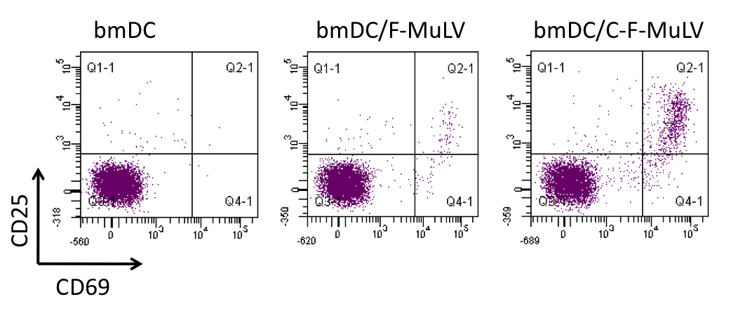
Figure 1. Complement-mediated enhancement of the activation of specific CD8 T cells by bone marrow-derived DCs (bmDC) loaded with non-opsonized (F-MuLV) vs. complement-opsonized (C-F-MuLV) Friend Virus. Activation was measured by the coexpression of CD69 and CD25 on specific CD8 T cells.
II.) Interplay of complement and dendritic cells with oncolytic VSV-GP virotherapy
Oncolytic viruses (OV) represent a promising cancer therapy option by specifically replicating in and killing cancer cells while leaving healthy cells undamaged. The oncolytic virus, VSV-GP was developed by Professor von Laer and has been successfully tested in several xenograft and syngeneic models. Since oncolytic activity of VSV-GP might release antigens related to the cancer cells we are interested how VSV-GP oncolytic therapy influence dendritic cells functions and dendritic cell immunotherapy. Furthermore, a potential activation of the complement system by VSV-GP virotheraphy might i) limit oncolytic activity due to complement-mediated lysis of viral particles and ii) generate complement components potentially able to modify specific immune responses particularly through the modulation of DC functions. Thus we are aiming to understand this complex interplay between the complement system, DCs and the oncolytic virotherapy (Figure 2).
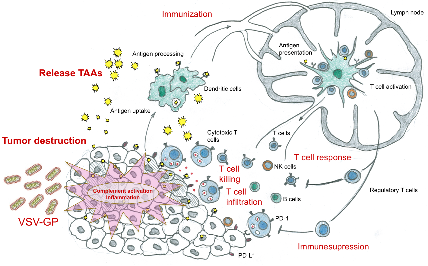
Figure 2. Interplay of complement and dendritic cells with oncolytic VSV-GP virotherapy (modified from Mellmann et al., 2011, Nature, 480:480)
III.) MAIT cell biology:
Mucosal associated invariant T (MAIT) cells expressing an invariant T cell receptor represent an innate-like T cell population involved in immune defence against bacteria. In humans MAIT cells comprise about 10% of the CD8+ T cells and are enriched in mucosal tissues and the liver. MHC-related protein 1 (MR1) expressed on antigen presenting cells has been shown to present vitamin B metabolites synthesized by bacteria but not mammals. Riboflavin-metabolizing bacteria have been demonstrated to activate MAIT cells (Figure 3). Since in normal human serum (NHS) bacteria might activate the complement system, we investigate the modulation of bacteria-induced MAIT cell activation in NHS. This research topic is performed in close collaboration with Prof. Klenerman at the University of Oxford, UK.
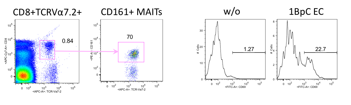
Figure 3. CD8+ TCRVa7.2+ CD161++ MAIT cells (dot plots) are activated in PBMCs in the presence of 1 bacteria per cell (BpC) Escherichia coli (EC) (histograms, w/o vs. 1BpC EC).
Group members:
- Mag. Brigitte Müllauer, Technician
- Jacqueline Schörgenhuber, M.Sc., Technician
- Jasmin Hatami, M.Sc., PhD-student
Former members:
- Dominik Klaver
- Simon Kruis
- Daniela Krenn
- Iris Koske
- Muneeb Idrees
- Nita Venkatesh