
RESCH microscopy
RESCH stands for “refocusing after scanning using helical phase engineering” and can be considered a variant of Image Scanning Microscopy where the imaging PSF has been changed such that 3D information from the specimen can be efficiently collected. One possibility is to shape the detection PSF of a scanning microscope into a helical shape as shown in Fig. 2. Suitable data post-processing allows for the reconstruction of volumetric data from a single planar scan.
 |
|
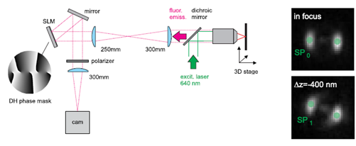
Figure 1: Optical setup for RESCH microscopy. A helical phase mask (here a mask encoding a double-helix) in the emission path of an Image Scanning Microscope allows for retrieving 3D data from 2D scans. |
 |
|
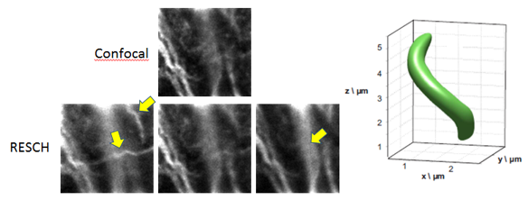
Figure 2: Left: Experimental results from the imaging of Alexa 647 stained microtubules in fixed COS 7 cells (NA 1.35, wavelengths: ex./em.=640/670 nm). The upper image shows a confocal image. The lower three are RESCH images of the same area which were all constructed from the same data set collected in a single 2D scan. |
Since RESCH collects a small volumetric slice in a single 2D scan, the application of 3D deconvolution tools to RESCH data already provides high axial resolution without the need to record z-stacks. An experimental result is shown in figure 3.
Also the lateral resolution is improved to a value better than provided by confocal microscopy, because the multi-view deconvolution approach inherently implements “pixel reassignment” as used in ISM.
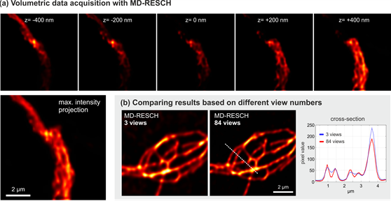 |
| Figure 3: Experimental RESCH data, collected in a single 2D-scan and post-processed with a maximum-likelihood multi-view deconvolution algorithm. Note the high optical sectioning (NA 1.25, wavelength: ex./em.=640/670 nm). |
Collaborations
Rainer Heintzmann, Leibniz Institute of Photonic Technology, Albert-Einstein-Strasse 9, Jena D-07745, Germany
Rafael Piestun, Department of Electrical and Computer Engineering, University of Colorado, Boulder, CO 80309, USA
Publications
Jesacher, A., M. Ritsch-Marte, and R. Piestun. 2015. Three-dimensional information from two-dimensional scans: a scanning microscope with postacquisition refocusing capability. Optica 2:210-213.
Roider, C., R. Heintzmann, R. Piestun, and A. Jesacher. 2016. Deconvolution approach for 3D scanning microscopy with helical phase engineering. Opt. Express 24:15456-15467.