
Optical nanoscopy by single molecule localization
Single-molecule localization microscopy (SMLM) denotes a family of methods to obtain optical “super-resolution” of fluorescently labelled objects down to the nanometer range. The concept is to resolve objects in time rather than space: Many recordings are taken, each containing only relatively few, sparsely scattered and spatially isolated molecules. This prevents the requirement to spatially resolve them. Instead, they are individually localized, which can be done with a much higher precision compared to the classical Abbe limit.
We investigate methods to obtain optimal localization performance for given experimental boundary conditions. The focus lies on obtaining highest localization precision whilst minimizing systematic errors that are very often unavoidable in SMLM.
In a collaboration with the group of Prof. Gerhard Schütz (TU Vienna), we concentrated on the presence of a nearby glass medium (typically the glass coverslip which separates objective lens from specimen), which can boost the localization precision along the optical axis by a factor of about 2 due to a near-field coupling effect.
We could show that this effect can be efficiently exploited by imaging at a small intentional defocus [2]. In following research we investigated the potential of popular 3D SMLM strategies (biplane or cylindric lens imaging) to “harvest” information contained in the near-field [3].
  |
|
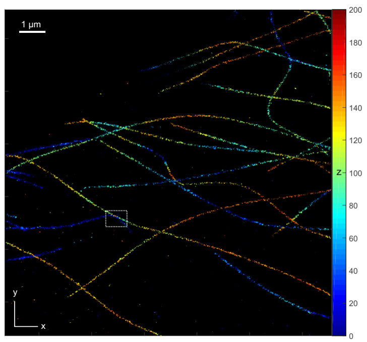
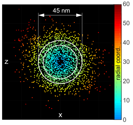
Figure 1: Left: SMLM image of Alexa 647 stained microtubuli in a COS-7 cell. The z-coordinate is color coded (scale in nm). Right: Cross section of an averaged microtubule, revealing its hollow core. The raw data for these images has been acquired using off-focus SMLM [2] using a high-NA objective lens (NA = 1.7). |
Researchers on this project at our institute
- Julian Maloberti, PhD
- Alexander Jesacher, PhD (PI)
Funding
“3D nanoscopy of the immunological synapse” FWF P30214, 2017 – 2021.
„Live Cell Superresolution Imaging of Protein Conformation“, FWF P-36022B, 2022 – 2026
Collaborations
- Gerhard Schütz, Inst. Of Applied Physics, TU Vienna
- Martin Offterdinger, Division of Neurobiochemistry, Medical University of Innsbruck
Publications
[1] P. Zelger, K. Kaser, B. Rossboth, L. Velas, G. J. Schütz, and A. Jesacher, „Three-dimensional localization microscopy using deep learning,“ Opt. Express 26, 33166-33179 (2018)
[2] Philipp Zelger, Lisa Bodner, Lukas Velas, Gerhard J. Schütz, and Alexander Jesacher, „Defocused imaging exploits supercritical-angle fluorescence emission for precise axial single molecule localization microscopy,“ Biomed. Opt. Express 11, 775-790 (2020)
[3] Philipp Zelger, Lisa Bodner, Martin Offterdinger, Lukas Velas, Gerhard J. Schütz, and Alexander Jesacher, „Three-dimensional single molecule localization close to the coverslip: a comparison of methods exploiting supercritical angle fluorescence,“ Biomed. Opt. Express 12, 802-822 (2021)
[4] Philipp Zelger, PhD thesis “Three-dimensional single molecule localization close to the coverslip”, 2020.
Software download
Here you can download our Matlab apps for evaluating SMLM data taken in TIRF configuration: link
Department für Physiologie und Medizinische Physik
- Department für Physiologie und Medizinische Physik
- Institut für Physiologie

- Forschungsgruppen
- M. Kress
- Team
- Publications
- Techniques
- Gallery
- B. Flucher
- Team
- Molecular Membrane Physiology & Functional Proteomics
- Cell and Tissue Culture
- Mitarbeiter:innen und Kontakt
- Öffentliche Ressourcen
- Jobs
- Institut für Biomedizinische Physik
- Department für Physiologie und Medizinische Physik
- Institut für Physiologie
- Forschungsgruppen
- M. Kress
- Team
- Publications
- Techniques
- Gallery
- B. Flucher
- Team
- Molecular Membrane Physiology & Functional Proteomics
- Cell and Tissue Culture
- Mitarbeiter:innen und Kontakt
- Öffentliche Ressourcen
- Jobs
- Institut für Biomedizinische Physik