
Research Groups
UV Radiation
Group Leader: Axel Kreuter
|
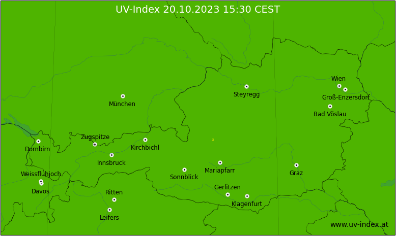
Current UV-Index in Austria |
We are interested in various aspects of solar radiation with emphasis on the UV spectral range. We develop new spectroradiometric instruments and analysis techniques and measure solar radiation spectra and aerosol optical depth in Innsbruck and during dedicated campaigns. We also operate the Austrian UV measurement network. see also: UV-Index |
Biomedical Laser Applications
Group Leaders: Monika Ritsch-Marte, Stefan Bernet, Alexander Jesacher, and Gregor Thalhammer-Thurner
Optical Micromanipulation
|
Optical micromanipulation stands for contact-free handling of microscopic particles with laser light. With „Optical Tweezers“ one can handle microscopic particles such as micro-organisms, micro-beads, living cells, cell organelles, or DNA-strands. Our group has developed methods to shape optical trapping patterns with computer-holography methods. Moreover, we strive at pushing the limits of trapping towards increasingly large particles, including fast swimming organisms such as flagellates or Euglena species. read more |

SLM-aided optical imaging:
|
We use miniaturized liquid crystal displays (LCDs) with individually addressable micrometer-sized pixels, so-called Spatial Light Modulators (SLMs), to advance optical microscopy. SLMs can emulate contrast mechanisms (e.g. brightfield, darkfield or phase contrast), create multiplexed images (combining different imaging settings in one recorded image), and steer or pattern the illumination in the microscope. read more |
Adaptive multi-photon imaging
|
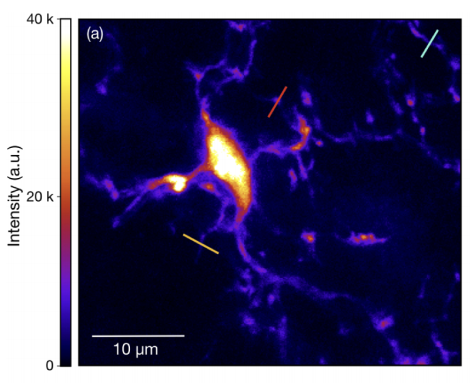
Multi-photon excitation scanning microscopy such as two-photon excitation fluorescence (TPEF) is one of the most powerful tools for imaging structures deep in biological tissues. We use adaptive optics to compensate the scattering induced by tissue and therefore to increase the imaging depth. read more |
Optical nanoscopy by single molecule localization
|
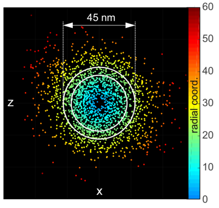
Single-molecule localization microscopy (SMLM) denotes a family of methods to obtain optical “super-resolution” of fluorescently labelled objects down to the nanometer range. The concept is to resolve objects in time rather than space. We develop methods to improve on localization precision and robustness for specific experimental boundary conditions. read more |
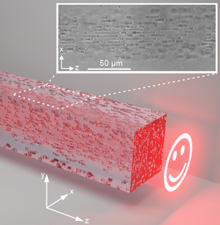
3D diffractive elements through fs-laser direct writing
|
Direct laser writing using ultra-short pulsed lasers is an increasingly used technology for the generation of embedded photonic structures such as waveguides or holographic data storage. One underlying fundamental process is the controlled refractive index change of transparent dielectrics by laser irradiation. We work on numerical tools for the design of such structures and experimental methods for their optical characterization. read more |
Former projects
Chemical imaging with Coherent anti-Stokes Raman Scattering (CARS)
|
If the frequency difference of two laser beams matches a Raman-active vibration of targeted molecules, a blue-shifted anti-Stokes signal is generated. This effect can be exploited for spectroscopy. Due to its coherent nature, CARS can lead to much higher signal strengths than e.g. Raman spectroscopy. The combination of CARS with microscopic imaging leads to CARS microscopy, which enables fast recording of chemical maps on micrometer scale. From a practical point of view, CARS microscopy resembles fluorescence microscopy, but without the need for fluorescent dyes. We have developed a non-scanning CARS microscope where an extended area in the sample plane is simultaneously illuminated by pump and Stokes beams under phase matching conditions. |
More information:
Department für Physiologie und Medizinische Physik
- Department für Physiologie und Medizinische Physik
- Institut für Physiologie

- Forschungsgruppen
- M. Kress
- Team
- Publications
- Techniques
- Gallery
- B. Flucher
- Team
- Molecular Membrane Physiology & Functional Proteomics
- Cell and Tissue Culture
- Mitarbeiter:innen und Kontakt
- Öffentliche Ressourcen
- Jobs
- Institut für Biomedizinische Physik
- Department für Physiologie und Medizinische Physik
- Institut für Physiologie
- Forschungsgruppen
- M. Kress
- Team
- Publications
- Techniques
- Gallery
- B. Flucher
- Team
- Molecular Membrane Physiology & Functional Proteomics
- Cell and Tissue Culture
- Mitarbeiter:innen und Kontakt
- Öffentliche Ressourcen
- Jobs
- Institut für Biomedizinische Physik
Contact
Director
o. Univ.-Prof. Dr. Monika Ritsch-Marte
Secretary
Michaela Palz
Phone: +43 (0)512/9003-70871
Fax: +43 (0)512/9003-73871
Mail: office.biomedphys@i-med.ac.at
Müllerstraße 44
A-6020 Innsbruck