
Investigating the single-cell metabolism of lipids
We demonstrated the ability to differentiate between various vegetable oils at the signatures of the weak =C-H stretching vibration around 3015cm-1 and obtained quantitative spectra that measure the ratio between saturated and unsaturated fatty acids in vegetable oils. This allows investigating the lipid metabolism of adipocytes on a single cell level.
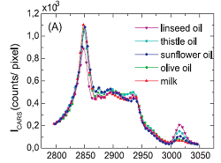 |
Fig. 1 shows CARS spectra representing the typical signatures of lipids in the Raman shift region between 2750 and 3050cm-1. They result from a superposition of several different vibrational states of symmetric and asymmetric CH2 and CH3 stretching vibrations. The resonance at 3015cm-1 depends on the number of C=C double bonds, since it corresponds to the C-H stretching vibration of a hydrogen attached to a C=C group and therefore varies strongly with the degree of saturation of vegetable oils.
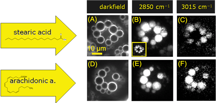 |
Our CARS set-up was used to image lipids within adipocyte cell cultures. Different populations of the same cell line had previously been fed on various saturated and unsaturated fatty acids. This "diet" led to different average concentrations of 5.8% and 9.8% of polyunsaturated fatty acids in the internal fat vesicles (measured with HPLC). The cells were imaged with CARS at the strong resonance of the saturated -C-H bindings at 2850cm-1 and at the weak peak of the unsaturated =C-H bindings at 3015cm-1. The results are shown in Fig. 2. The ratios between the weak =C-H and the strong -C-H peaks were measured to be 1.7% and 3.3% for the two cell lines fed of stearic and arachidonic acid. The approximately 4% difference in the concentration of polyunsaturated fatty lipids could be clearly detected.
Publication(s):
C. Heinrich, A. Hofer, A. Ritsch, C. Ciardi, S. Bernet, and M. Ritsch-Marte, Selective imaging of saturated and unsaturated lipids by wide-field CARS-microscopy, Opt. Express, 16, 2699 (2008).