
Pathophysiology
Research Focus
General Facts
Research
Selected Publications
Selected Funding, Collaboration
Keywords: Mitosis, Cell Cycle Regulation, Tumor, Kinase, Fibrosis, Systemic sclerosis, Endoplasmic reticulum, Golgi apparatus, Proteostasis, Membrane Trafficking
Research (ÖSTAT Classification): 106023, 106052, 106002, 301109, 301108, 301105, 301101, 301203, 301306, 301904, 304003, 301902
Research Focus
Our work focuses on mechanisms that contribute to tumorigenesis and systemic sclerosis. We use cell-based systems, primary patient material and model organisms to try to address questions concerning the regulation of cell death in leukaemia, the regulation of mitosis and fidelity of chromosome segregation, the characterisation of CCNY-dependent kinases, the role of RNA-binding proteins in gene expression required for proliferation, and mechanisms in systemic sclerosis.
General Facts
The Institute of Pathophysiology has a longstanding interest in the molecular mechanisms that control cellular proliferation and cell death in pathophysiological conditions such as cancer and in understanding the pathophysiology of chronic diseases such as systemic sclerosis.
We use interdisciplinary approaches and collaborate at both national and international level for better understanding of the mechanisms of chromosome congression and segregation during mitosis as well as the role of the mitotic ubiquitin ligase APC/C, in order to reveal mechanisms that might contribute to aneuploidy, a common hallmark of human malignancies.
Beyond mitosis, we focus on cell-cycle-unrelated functions of cyclin-dependent kinases and try to understand the role of RNA-binding proteins in the control of gene expression, which may be relevant for proliferation.
Based on our analysis of systemic sclerosis in an animal model, we proposed that systemic sclerosis is an autoimmune disease directed against microvascular endothelial cells and we are now testing approaches to translate this knowledge into therapy.
The Laboratory of Autoimmunity (LAI), established in 2007 and led by em.o.Univ.-Prof. Georg Wick, closed in 2018. The work of the LAI focused on two topics: the immunology of fibrosis and the immunology of atherosclerosis.
In December 2020, Prof. Hesso Farhan was appointed as the new head of the Institute of Pathophysiology. His research group is interested in the following topics:
- Regulation of membrane trafficking by kinase signaling
- Mechanisms how cells maintain their protein balance (proteostasis) and how de-regulated proteostasis is linked to diseases such as neurodegeneration and multiple myeloma
- Characterize the role of cell migration of breast cancer progression
- Role of pseudoenzymes in physiology and pathology.
Research
Systemic Sclerosis
Roswitha Sgonc
Our group is interested in the pathogenesis and treatment of systemic sclerosis (SSc), which is studied in human patients as well as in the spontaneous avian model UCD-200/206, the only animal model that manifests the complete clinical, histopathological and serological spectrum of human SSc. This comparative study of UCD-200/206 chicken and human SSc made it possible to identify microvascular endothelial cells as the primary target of the autoimmune attack. After many years studying pathomechanisms and genetic factors underlying the disease, we are now focusing on the development of novel therapeutic approaches.
There is an unmet need for effective pro-angiogenic treatment of ischaemic lesions in patients with SSc. Vascular alterations in both human and avian SSc predominantly affect the microvasculature. Tissue hypoxia normally induces angiogenesis but vascular repair and angiogenesis seem to be strongly disturbed with SSc, owing to dysregulation of vascular endothelial growth factor (VEGF). In our therapeutic attempts, we aim to manipulate VEGF levels by using a VEGF121 variant that covalently binds to fibrin and is released only on demand by cellular enzymatic activity. We have shown that cell-demanded release of locally applied fibrin-bound VEGF121 leads to the formation of morphologically normal blood vessels as well as clinical improvement of early and late ischaemic lesions. Long-term studies showed lasting effects on the improvement and prevention of ischaemic lesions and these support the notion that cell-demanded release of fibrin-bound VEGF is capable of translating a supra-physiological dose into a physiological tiny dose and of sustaining this dose for long enough to allow vessels to mature into stable vessels.
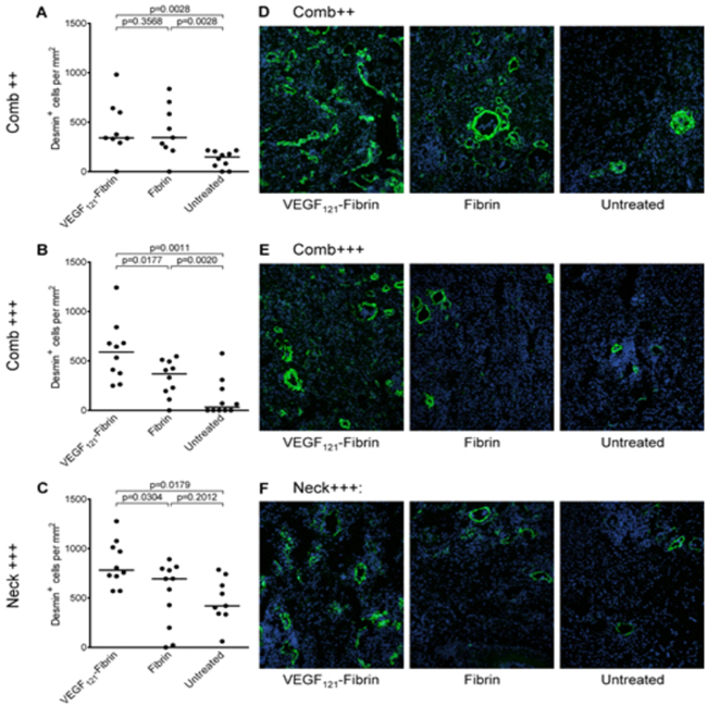
Fig 1: VEGF121-fibrin treatment of ischaemic lesions leads to growth of stable blood vessels. Desmin-stained mural cells were quantified on immunofluorescence-stained frozen tissue sections after one week of treatment of early inflammatory comb lesions (C++; A), comb ulcers (C+++; B) and neck ulcers (N+++; C). P values were calculated using the Mann-Whitney-U test adjusted by the Kruskal-Wallis test. Each dot represents a single lesion. Horizontal bars indicate median values. Representative false colour overlay pictures: desmin and DAPI staining (D-F). Original magnification: 200x.
Cell Cycle Control
Stephan Geley
Cyclin-dependent kinases (CDKs) are proline-directed serine threonine kinases, which consist of a catalytic and a regulatory subunit, the cyclin. CDKs comprise a family of 20 different cyclin-CDK pairs that are best known for their role in cell cycle regulation and transcription. The cell cycle regulator CDKs are negatively regulated by the mitotic ubiquitin ligase APC/C, which targets the mitotic cyclins for proteasome-dependent degradation. APC/C activity during mitosis requires CDK activity and the WD40 repeat protein CDC20, and these are required for the onset of anaphase and for exit from mitosis. In the ensuing G1 phase, CDC20 is substituted with CDH1/FZR1, to keep the APC/C active until the onset of DNA replication. The switch from high CDK activity during mitosis to low CDK activity during the G1 phase is required in order to allow resetting of the cell division cycle, for replication licensing, exit from the cell division cycle, initiation of differentiation and ciliogenesis.
The onset of anaphase and the activation of the APC/C during mitosis are regulated by the spindle assembly checkpoint (SAC). Non or wrong interactions of kinetochores with dynamic microtubules of the mitotic spindle generate an inhibitory signal, the mitotic checkpoint complex, MCC, which binds to and inactivates CDC20 to keep the APC/C inactive until correct bipolar attachment to the mitotic spindle is achieved. This inhibition is relieved only on bipolar attachment of the kinetochores to the microtubules, which produces mechanical tension within and between the kinetochores, resulting in SAC silencing. Bipolar chromosome attachment and the congression of chromosomes to the metaphase plate require several factors, including chromokinesins, plus-end directed microtubule-dependent motor proteins that bind to chromosomes. We showed that chromokinesins are required for chromosome congression to the metaphase plate and that they are involved in regulating microtubule dynamics.
SAC activity can be maintained if SAC silencing factors are reduced, and one of these factors is the dynein motor protein-dependent removal of kinetochore SAC signalling proteins. Spindly is a small dynein adaptor that is necessary for dynein localisation to the kinetochore and that requires protein farnesylation for its functions. To avoid mislocalisation of Spindly and therefore dynein to cellular membranes as a result of increased lipophilicity, the farnesylation of Spindly is tightly regulated.
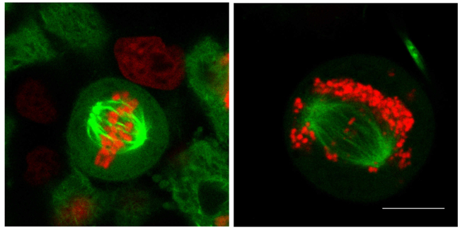
Fig. 2: Mitosis without chromosome attachment. Right panel: Control mitotic HeLa cells expressing histone H2B-RFP and a-tubulin-GFP. Left panel: After disruption of kinetochores, cells were allowed to enter mitosis. Cells arrest owing to the activation of the SAC. Confocal images taken on a Leica SP5 confocal microscope. Scale bar: 5µm
Cell cycle regulation depends on the execution of a tightly controlled gene expression programme, controlled by regulated transcription, translation, post-translational modification and proteolysis. RBM26 is a poorly characterised protein that is required for proliferation in Drosophila. In human cells, RBM26 is an inessential gene but, together with its homologue RBM27, it controls the turnover of long non-coding RNAs, an essential function in human cells.
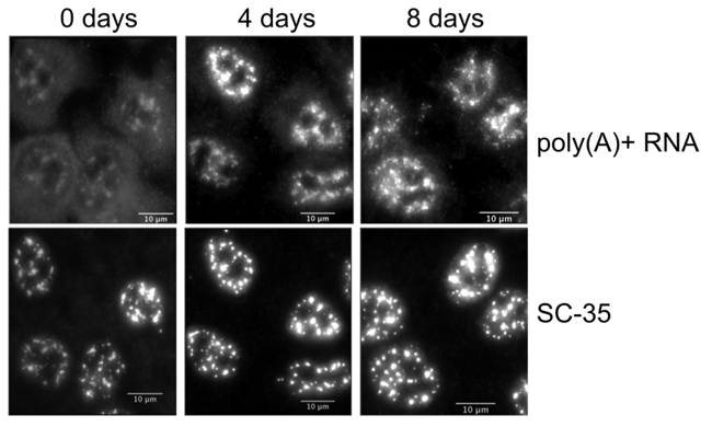
Fig. 3: RNA binding proteins RBM26 and 27 are required for RNA homeostasis. Depletion of RBM27 was induced over 8 days in a cell line that lacked RBM26 gene expression. Cells were stained for poly(A+)-RNA by means of in-situ hybridisation using a Cy3-labelled oligo-dT probe and the nuclear speckle marker SC-35. Upon depletion of these RBM proteins, poly(A)+ RNA accumulates in the nucleus and causes accumulation of SC-35.
After exit from mitosis, the APC/C remains active to retrain premature entry into the S-phase. In addition, downregulation of mitotic kinase activity is required, in order to establish an environment that allows ciliogenesis. Loss of FZR1/CDH1 maintains a low level of mitotic kinase activity and thus impairs ciliogenesis, which might explain the developmental phenotypes observed in FZR1/CDH1-deficient mice.
Cyclin-dependent kinases are involved not only in cell cycle control but also in transcription, regulation of the cytoskeleton and more. CDK16 is a poorly characterised kinase that is activated by membrane-bound cyclin Y (CCNY). CDK16 belongs to a group of related kinases that can all be activated by CCNY and are therefore likely to act redundantly; this impedes functional characterisation of these kinases. We therefore used in vitro substrate screening to define a kinase substrate consensus motif, which we employed to define potential substrates including the microtubule-binding protein doublecortin. In addition, we demonstrated that, when expressed together with CCNY, the recombinant active kinase also contains 14-3-3 proteins and we showed that these accessory proteins are required for kinase activity.
Selected Publications
Research Group Wick
- Jakic B., Carlsson M., Buszko M., Cappellano G., Ploner Ch., Onestingel E., Foti M., Hackl H., Demetz E., Dietrich H., Wick C., Wick G.: The Effects of Endurance Exiercise and Diet on Atherosclerosis in Young and Aged ApoE-/- and Wild-Type Mice. Gerontology 30:1-12. doi: 10.1159/000492571 (2018)
- Jakic B., Wick G., Cappellano G.: Hsp60 in Atherosclerosis: Past, Present and Future. In: Asea A., Kaur P. (eds) Heat Shock Protein 60 in Human Diseases and Disorders. Heat Shock Proteins, vol 18. Springer, Cham (2019)
- Jakic B., Kerjaschki D., Wick G.: Lymphatic Capillaries in Aging. Gerontology 2020, DOI: 10.1159/000508459
Research Group Sgonc
- Allipour Birgani S, Mailänder M, Wasle I, Dietrich H, Gruber J, Distler O, Sgonc R (2016): Efficient therapy of ischaemic lesions with VEGF121-fibrin in an animal model of systemic sclerosis. Ann Rheum Dis. 75:1399-406.
Research Group Geley
- Shehata SN, Deak M, Collodet C, Spiegl SF, Geley S, Sumpton D, Sakamoto K.: Identification of novel PCTAIRE-1/CDK16 substrates using a chemical genetic screen. Cell Signal. 2019 Jul;59:53-61. doi: 10.1016/j.cellsig.2019.03.012. Epub 2019 Mar 14.
- Dohmen M, Krieg S, Agalaridis G, Zhu X, Shehata SN, Pfeiffenberger E, Amelang J, Bütepage M, Buerova E, Pfaff CM, Chanda D, Geley S, Preisinger C, Sakamoto K, Lüscher B, Neumann D, Vervoorts J.: AMPK-dependent activation of the Cyclin Y/CDK16 complex controls autophagy. Nat Commun. 2020 Feb 25;11(1):1032. doi:10.1038/s41467-020-14812-0.
- Steblyanko Y, Rajendraprasad G, Osswald M, Eibes S, Jacome A, Geley S, Pereira AJ, Maiato H, Barisic M.: Microtubule poleward flux in human cells is driven by the coordinated action of four kinesins. EMBO J. 2020 Oct 19:e105432. doi: 10.15252/embj.2020105432. Epub ahead of print.
Selection of Funding
Collaborations
- Oliver Distler, Department of Rheumatology, Center of Experimental Rheumatology, University Hospital Zurich, Zurich, Switzerland
- Marin Barisic, Danish Cancer Center, Copenhagen, Denmark
- Kai Sakamoto, Novo Nordisk Foundation, Copenhagen, Denmark
- Jörg Vervoorts, RWTH Aachen University, Aachen, Germany
 ao.Univ.-Prof. Dr.med.univ. Stephan Geley
Director (until December 2020)
Contact:
Innrain 80-82
6020 Innsbruck
Austria
Email: stephan.geley@i-med.ac.at
Phone: +43 512 9003 70365
Fax: +43 512 9003 73100
https://biocenter.i-med.ac.at/pathophysiology/
 Univ.-Prof. Dr.med.univ. Hesso Farhan
Director (since December 2020)
Contact:
Innrain 80-82
6020 Innsbruck
Austria
Email: hesso.farhan@i-med.ac.at
Phone: +43 512 9003 70360
Fax: +43 512 9003 73100
https://www.i-med.ac.at/pathophysiologie/