search_icon 

close_icon

search_icon  

search_icon  

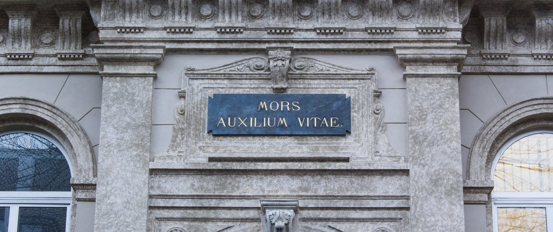
Informationen für Studierende

Ohne profundes morphologisches Wissen sind Entstehung und Entwicklung von Krankheiten des Nervensystems nicht zu begreifen. Die Neuroanatomie vermittelt Kenntnisse von der Gestalt und Feinstruktur des zentralen und peripheren Nervensystems und ist somit die Basis ärztlichen Handelns in den Neuro-Fächern. 

An der MUI bekommen Sie eine praktische Ausbildung an anatomischen und mikroskopischen Präparaten, die notwendig ist, um ärztlichen Fehlern bei Diagnose und Therapie vorzubeugen. Außerdem werden zelluläre und molekulare Grundlagen des Nervensystems vermittelt, die wesentlich zum Verständnis von Neurologie und Psychiatrie beitragen. Im Mittelpunkt des Unterrichts steht aber die funktionelle Neuroanatomie. Sie bildet einen wesentlichen Teil der Neurobiologie des Menschen - einer Wissenschaft für angehende Mediziner, die lieber verstehen als auswendig lernen.

Die Neuroanatomie-Vorlesung umfasst Zeichenvorlagen und Schemata zur Verdeutlichung der Lehrinhalte, die den ‚Nomina Anatomica’ und dem ‚Gegenstandskatalog für die vorklinische Medizin’ (Mainz, 1999) entnommen sind. Für das ZNS-Praktikum gilt, dass anatomische und histologische Begriffe an einem Beispiel erklärt und am Präparat zu demonstrieren sind. Umgekehrt sollen sichtbare Strukturen benannt werden. Dazu gehören auch die Blutgefäße und das Ventrikelsystem von Gehirn und Rückenmark.

Wir verwenden neben makroskopischen Originalpräparaten (im Rahmen des Sezierkurses) und Plastinaten eine Vielzahl von Gewebeschnitten, die am Lichtmikroskop und digital zu beurteilen sind, sowie Schnittbilder aus unserer neuroradiologischen Klinik und von IMAIOS. Grundlegende neuroanatomische und neurohistologische Arbeitsmethoden sollten ebenso beherrscht werden.

Die im Corona-Wintersemester 2020/21 aufgezeichneten Vorlesungen sind im E-Learning-Programm der MUI (MOODLE) abrufbar. Begleitend zur Vorlesung und für die Prüfungsvorbereitung bietet sich für die Neuroanatomie außerdem ein Kompendium des Institutsleiters an, das hier heruntergeladen werden kann. Die zur Verfügung gestellten Lernunterlagen erlauben es, die für die weitere klinische Ausbildung notwendigen Grundlagen aus Lehrbüchern zu extrahieren, keinesfalls können sie das Lehrbuchstudium oder den Vorlesungsbesuch ersetzen.

Wir empfehlen insbesondere das Buch Neuroanatomie: Nachschlagen, Lernen, Verstehen von Markus Kipp und Kalinka Radlanski (KVM, 3. Auflage 2022), da es am Prüfungswissen orientiert ist und die Lernziele des Unterrichts an der MUI nahezu vollständig abbildet. Die histologischen Aspekte der Neuroanatomie werden in einem Skriptum von Hans-Georg Frank und Katharina Sternecker behandelt, das auch der bei uns gelehrten digitalen Histologie zugrunde liegt. Genaue Lokalisationen von neuronalen Kerngebieten und Bahnen sind im Taschenatlas 'Das Zentralnervensystem des Menschen' von Rudolf Nieuwenhuys und Kollegen nachzuschauen. Neurowissenschaftlich besonders Interessierten wird das Standardwerk von Mark Bear et al. empfohlen (Springer Spektrum, 4. Auflage 2018).

Für eine weitergehende Lektüre zur Hirnentwicklung, zum Abbau unseres Gehirns im Alter und zu neurodegenerativen Erkrankungen empfehlen wir ein Sachbuch des Institutsleiters, das 2021 im Springer-Verlag erschienen ist.

----------------------------------------------------------------------------

Information for students

Without profound morphological knowledge, the origin and development of diseases of the nervous system cannot be understood. Neuroanatomy provides knowledge of the shape and fine structure of the central and peripheral nervous system and is thus the basis of medical action in the neuro-subjects.
 
At MUI, you will receive hands-on training on anatomical and microscopic specimens, which is necessary to prevent medical errors in diagnosis and therapy. You will also learn cellular and molecular principles of the nervous system, which are essential to understanding neurology and psychiatry. However, functional neuroanatomy is the focus of our teaching. It forms an essential part of human neurobiology - a science for aspiring physicians who prefer to understand rather than memorize.
 
The neuroanatomy lecture includes drawing templates and diagrams to clarify the teaching content, which are taken from the 'Nomina Anatomica' and the 'Gegenstandskatalog für die vorklinische Medizin' (Mainz, 1999). For the CNS practical course, anatomical and histological terms are to be explained by example and demonstrated on the specimen. Conversely, visible structures are to be named. This includes blood vessels and the ventricular system of the brain and spinal cord.
 
We will use macroscopic original specimens (as part of the dissection course) and plastinates, as well as a variety of tissue sections to be assessed on the light microscope and digitally, and cross-sectional images from our neuroradiology clinic and IMAIOS. Basic neuroanatomical and neurohistological working methods should also be mastered.
 
Lectures recorded in the Corona winter semester 2020/21 are available in the MUI e-learning program (MOODLE). Accompanying the lecture and for exam preparation, a Compendium by the Institute Director is also available for Neuroanatomy. The learning materials provided allow to extract the basics necessary for further clinical training from textbooks, in no way can they replace textbook study or lecture attendance.
 
In particular, we recommend the book Neuroanatomy: Reference, Learning, Understanding by Markus Kipp and Kalinka Radlanski (KVM, 2nd edition 2018), as it is oriented to exam knowledge and almost completely maps the learning objectives of the classes at MUI. The histological aspects of neuroanatomy are covered in a script by Hans-Georg Frank and Katharina Sternecker, which is also the basis for the digital histology taught at our institute. Precise localizations of neuronal nuclei and pathways can be found in the pocket atlas 'The Human Central Nervous System' by Rudolf Nieuwenhuys and colleagues. For those particularly interested in neuroscience, the standard work by Mark Bear et al. is recommended (Springer Spektrum, 4th edition 2018).
 
For further reading on brain development, the degradation of our brain in old age and neurodegenerative diseases, we recommend a non-fiction book by the director of the institute, published by Springer in 2021.

Institute of Neuroanatomy


Prof. Dr. med. Lars P. Klimaschewski

Medical University Innsbruck
Department of Anatomy & Histology
Institute of Neuroanatomy
Muellerstrasse 59
6020 Innsbruck, Austria
 
TEL +43 512 9003 71160
FAX +43 512 9003 73170
Email: lars.klimaschewski@i-med.ac.at


Prof. Dr. med. Lars P. Klimaschewski

Medical University Innsbruck
Department of Anatomy & Histology
Institute of Neuroanatomy
Muellerstrasse 59
6020 Innsbruck, Austria
 
TEL +43 512 9003 71160
FAX +43 512 9003 73170
Email: lars.klimaschewski@i-med.ac.at