Otorhinolaryngology
Research Focus
General Facts
Research
Selected Publications
Selected Funding, Collaboration
Keywords: Otorhinolaryngology, head & neck surgery; radioresistance; chemoresistance; oncolytic viruses; epithelial-mesenchymal transition; inner ear; hearing implants; neurobionic interface; therapeutic hypothermia; rhinosinusitis; computer aided surgery; radiomics; clinical outcomes research.
Research (ÖSTAT Classification): 302023, 302027, 302029, 302055
Research Focus
We focus on basic clinical and translational research in the fields of otology, rhinology, laryngology and head & neck cancer. Our goals are to improve diagnosis and clinical decision-making, to develop innovative treatment devices and surgical techniques, to advance precision medicine and to develop and validate instruments to measure treatment outcomes. We use high-throughput techniques, artificial intelligence, online databases and virtual reality on the basis of a large amount of experimental and clinical data.
General Facts
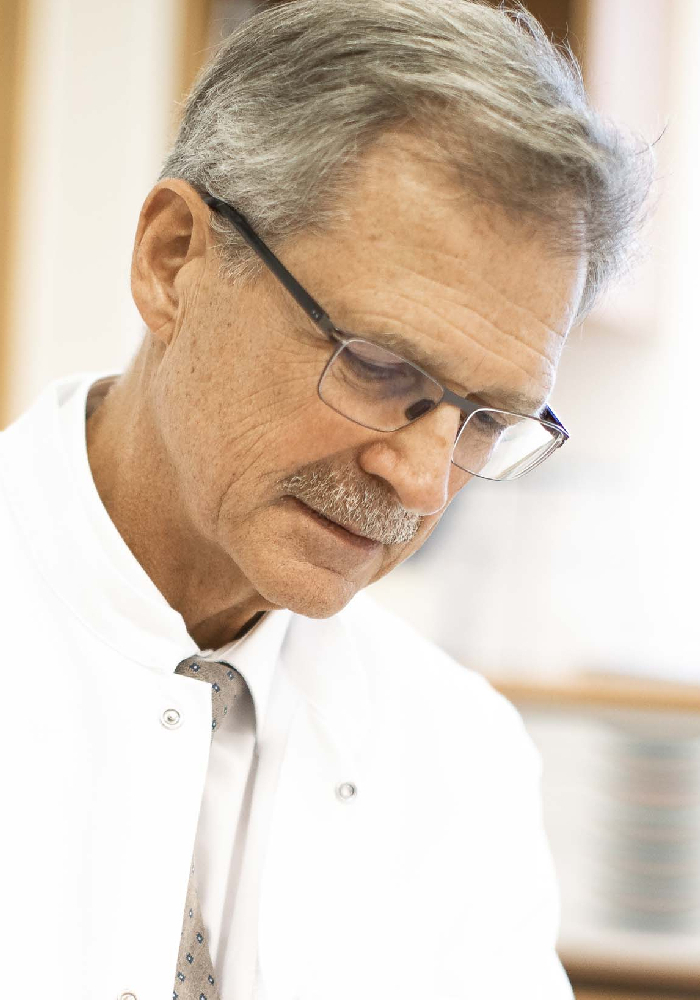
The University Hospital for Otorhinolaryngology provides healthcare for the greater Tyrolean area. It has 3 operating theatres, 3 wards with 41 beds and an outpatient clinic. On average, 2800 inpatients and 25,000 outpatients are treated here every year. In addition to all standard otorhinolaryngologic surgical procedures, we provide cochlear implants (Fig. 1), hypoglossal implants, sialendoscopy and Eustachian tube dilation. The state-of-the-art technical facilities include intraoperative navigation, neuromonitoring and various laser types such as CO2, KTP and Erbium laser.
We have 3 basic clinical research laboratories, a clinical head & neck cancer registry with over 1200 incident cases, a chronic rhinosinusitis patient registry (approx. 300 patients) and a scientific biobank (minus 190°C: 230 tumour samples, 100 paranasal sinus samples and 80 control tissues, i.e. healthy pharyngeal and nasal turbinate mucosa; FFPE: 800 tumour and 300 paranasal sinus samples; RNA isolates: 80 tumours and 12 controls [gene expression checked]).
Research
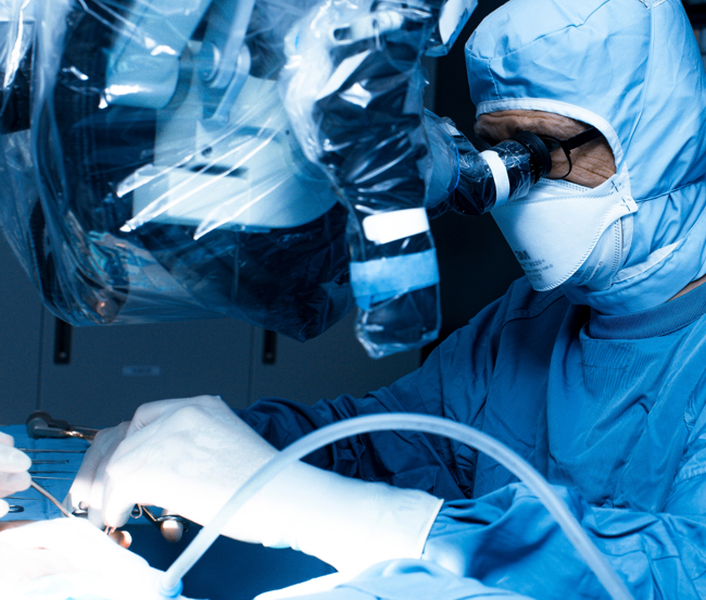
Based on micro-CTs of adult human temporal bones (middle ear, cochlea and vestibular organ) combined with high-resolution ultrastructural and structural data of tissue compartments, measurements of physiological parameters, gene and protein expression data, metabolic data and ion-channel distribution, we have developed a virtual 3D human temporal bone model (average of 10 Caucasian individuals) to optimise neurobionic device design and assess the effects of therapeutic hypothermia for inner ear protection during implant insertion (Fig. 2).
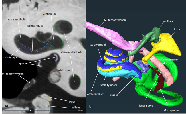
Similar methods were used to develop 3D patient-derived head & neck tumour culture systems for functional drug testing and therapy response prediction that is as realistic as possible. These models are being used in several projects, including efficiency testing of oncolytic viruses and the investigation of tumour/stroma interaction, tumour metabolism and epithelial-to-mesenchymal transition mechanisms (Fig. 3).
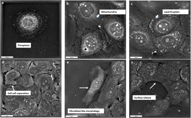
Multichannel immunofluorescence microscopy with automated image analysis of patient-derived tissues is used to characterise mechanisms of chronic rhinosinusitis and head & neck cancer, including identification of predictive biomarkers (Fig. 4). Machine learning and various proprietary algorithms for image analysis are also used for extraction of radiomic features in clinical imagery, surgical robotics and computer-aided surgical navigation.
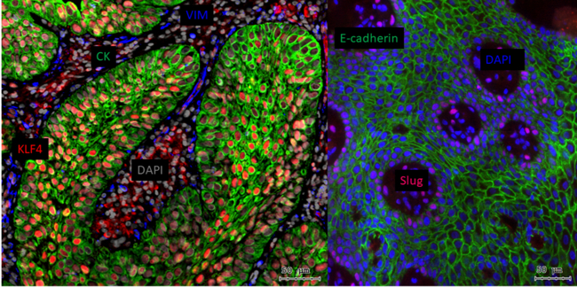
E-cadherin shows a decreasing gradient towards the periphery of the cancer cell nests. Slug-positive, dedifferentiated tumour cells are in epithelial-mesenchymal transition and E-cadherin negative.
Further research projects include peripheral arterial tonometry for the investigation of paediatric sleep apnoea, effects of head & neck cancer treatment on intestinal microbiome and cachexia, development and validation of instruments to assess clinical outcomes, and the role of viral infections in the pathogenesis of middle ear disease and head & neck cancer.
Current research projects include:
- 3D finite element models of the human inner ear: simulation & optimisation of electrical stimulation (FWF-DFG),
- Gene expression & mutation analysis platform to predict the success of therapy for cochlear implants (FWF/Japan Soc. for the Promotion of Science),
- Nerve fibre regeneration and integration of bioactive inner ear implants – fully integrated neurobionic interface (FWF-DFG-SNF),
- Applied vestibular implant development: prototype implantation and evaluation of targeting (MedEl, Uni. Maastricht, VetMed Vienna),
- Hypothermia for inner ear protection in hearing implants (MedEl),
- Correlative multimodal imaging in the inner ear – digital integration of Micro-CT high-throughput imaging of protein & gene expression,
- Inner ear cell and organ culture models,
- Radiomics and machine learning,
- Improvement of accuracy in skull base surgical navigation, Rhinospider,
- Robotic information-based cochlear implantation,
- Navigation-assisted brain stem implantation,
- Quantitative intraoperative quality assurance on the lateral skull base,
- Multichannel fluorescence microscopy with automated image analysis,
- 3D tumour patient-derived culture systems for functional drug testing and therapy response prediction,
- VSV oncolytic viruses,
- Metabolomics of head & neck cancer (Tyrolean Cancer Research Institute),
- Nitrogen tank storage and reactivation of patient-related expansive tissue culture (spheroid or 2D, planned Bridge project proposal with ViraT/Boehringer Ingelheim),
- Development and validation of instruments for the measurement of treatment outcomes,
- TCGA database analyses (in cooperation with the Institute of Bioinformatics),
- Mechanisms of radioresistance and chemoresistance in head & neck cancer,
- Pathomechanisms of chronic rhinosinusitis, characterisation of inflammatory cells and immune regulatory cells using multichannel immunofluorescence microscopy,
- Intestinal microbiome in radiochemotherapy of head & neck cancer
- Routine laboratory tests and survival in patients with head and neck cancer
- WatchPAT registration in children with adenotonsillar hyperplasia.
Selected Publications
- Lechner M, Schartinger VH, Steele CD, Nei WL, Ooft ML, Schreiber LM, et al. Somatostatin receptor 2 expression in nasopharyngeal cancer is induced by Epstein Barr virus infection: impact on prognosis, imaging and therapy. Nat Commun. 2021;12(1):117. IF 12,121. (IF 11,090)
- Steinbichler TB, Dudas J, Skvortsov S, Ganswindt U, Riechelmann H, Skvortsova, II. Therapy resistance mediated by exosomes. Molecular cancer. 2019;18(1):58. (IF 15,302)
- Steinbichler TB, Savic D, Dudas J, Kvitsaridze I, Skvortsov S, Riechelmann H, et al. Cancer stem cells and their unique role in metastatic spread. Seminars in cancer biology. 2019. (IF 11,090)
Selection of Funding
(Project manager, Third-party funder, Titel, Funds raised (total))
- PD. Dr. Dudas, FWF I LA NKFIH, Predictive markers of immune checkpoint inhibitor therapy, 47.848,00 €
- PD. Dr. Dudas, FWF/Japan Soc. for the Promotion of Science, Neurotrophins in developing inner ear and HNSCC, 254.657,00 €
- Ao Prof. Freysinger, FFG Bridge 1, NAV-ABI, 358.920,00 €
- Prof. Riechelmann, Viratherapeutics AUT, 3D Primärkulturen von humanen Plattenepithelkarzinomen der Kopf-, Halsregion als Modell zur Infektion und Wirksamkeit onkolytischer Viren, 140.202,00 €
- Ass. Prof. Schmutzhard, Med-El AUT, Cool electrode, 81.050,00 €
- Ao Prof. Schrott-Fischer, Auton. Provinz Bozen, The aging cochlea, 128.458,00 €
- Ao Prof. Schrott-Fischer, SAT EFRE K-Regio, EVITA, 198.036,00 €
- Ao Prof. Schrott-Fischer, Med-El AUT, EVITA II, 107.506,00 €
- PD. Glückert, Med-El AUT, Ion channels in the inner ear, 225.486,00 €
- PD. Glückert, Med-El AUT, Gapless man-machine interface, 46.847,00 €
Collaborations
- Nassir NABAB, Computer science, TU Munic, Germany
- Marco Caversaccio, Department of Otorhinolaryngology, University of Berne, Switzerland
- Ron Kikinis, Brigahm & Womens Hospital, Harvard, Boston, MA, USA
- Ilona Kovalszky, Semmelweis University Budapest, Inst. Pathology and Experimental Cancer Research Budapest, Hungary
- Andrea Ladanyi, Department of Surgical and Molecular Pathology, National Institute of Oncology, Budapest, Hungary
- Shinshu University School of Medicine, Matsumoto, Japan
- Richard A. Altschuler Auditory Anatomy Laboratory, Kresge Hearing Research Institute, Ann Arbor, MI USA
- Helge Rask-Andersen, Department of Surgical Sciences, Otorhinolaryngology and Head and Neck Surgery, University of Uppsala, Sweden
- Frank Rattay, Computational Neuroscience and Biomedical Engineering, Institute for Analysis and Scientific Computing, Vienna University of Technology, Austria
- Ilmari Pyykko, Hearing and Balance Research Unit, Tampere University School of Medicine,Tampere, Finland
- Tracey Newman, University of Southampton, UK
- Azel Zine, Bioengineering and Nanoscience Laboratory, Université de Montpellier, Montpellier, France
- Andrew K. Wise, The Bionics Institute, East Melbourne, Australia
- Eybalin Michel, Inserm U 254 Neurobiologie de l’Audition, Montpellier, France
- Stephan Handschuh, VetCore Facility for Research Imaging Unit, V University of Veterinary Medicine, Vienna, Austria
- Andreas Mehrle, Mechatronik, MCI, Innsbruck, Austria
- Christian Baumgartner, Karl Fritscher, Rainer Schubert, Biomedical Computer Science and Mechatronics, UMIT Hall, Austria
- Monika Petersson, ViraTherapeutics GmbH, Bundesstraße 27, 6063 Rum, Austria
- Michael Vogele CEO, iSYS Medizintechnik GmbH, Kitzbühel, Austria
- Gernot Kronreif CEO, ACMIT Manufacturing GmbH, Wr. Neustadt, Austria
- Ingeborg Hochmair, Carolyn Garnham, Daniel Sieber, MED-EL Elektromedizinische Geräte Gesellschaft m.b.H., Fürstenweg 77a, Innsbruck, Austria
Devices & Services
Devices:
- Surgical navigation wet lab: simulation of intraoperative computer aided navigation (Prof. Freysinger)
- TissueFAXS i PLUS, TissueGnostics, Vienna: multichannel immunofluorescence microscopy (PD Dr. Dudas) and StrataQuest Software;
- Confocal Microscope Zeiss LSM510M, Carl Zeiss AG, Oberkochen, Germany (PD Dr. Glückert)
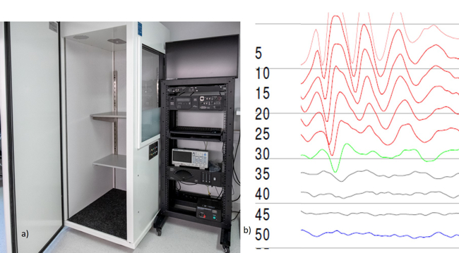
- Animal Auditory Brainstem Response Audiometry (Fig. 5), Tucker Davis, Alachua, Florida, USA: Registration of auditory brainstem potentials (hearing thresholds) in laboratory animals (Prof. Schrott-Fischer)
- JuLI BR Life Cell Analyzer, NanoEnTek Inc., Gyeonggi-do, Korea (PD Dr. Dudas)
- Infrared Laser Excitation for Quantitative Western Blot Imaging, Azure Biosystems, Dublin, CA, USA (PD Dr. Dudas)
Services:
- High speed intravital microscopy for diagnosis of ciliary dysfunction (PD Dr. Glückert)
- Ciliary ultrastructure for diagnosis of ciliary dysfunction (PD Dr. Glückert)
- Molecular characterization of cancer biopsies (PD Dr. Dudas)