
Biomedical Physics
Research Focus
General Facts
Research
Selected Publications
Selected Funding, Collaboration
Keywords: Biophotonics, digital holographic microscopy, super-resolution microscopy, spatial light modulators, optical tweezers, acousto-fluidics, linear and nonlinear Raman microscopy, diffractive optics, tunable lenses, UV radiation
Research (ÖSTAT Classification) : 206003, 103037, 302044, 103021, 103040
Research Focus
Innovative microscopy: Programmable SLM microscopy (wavefront shaping with spatial light modulators to emulate and customise “classic” microscopy techniques or to create new techniques), super-resolving single-molecule localisation microscopy, adaptive multi-photon excitation microscopy.
Highlights: Spiral phase contrast, multi-plane imaging, single-shot quantitative differential interference contrast, lensless imaging through a scattering medium, programmable confocal microscopy; axial super-localisation microscopy at the immunological synapse; high NA remote focusing methods.
Acoustic and optical micromanipulation: Contact-free handling of microscopic particles (micro-organisms, micro-beads, living cells, cell organelles or DNA-strands) with laser light.
Highlights: Trapping of the largest swimming micro-organisms ever trapped “all-optically”; combined acoustic and optical trapping of even larger specimens.
Solar UV radiation: Optimisation of spectroradiometric instruments and development of analysis techniques for solar radiation spectra and aerosol optical depth.
Highlights: Austrian UV measurement network (UV index).
General Facts
The Institute of Biomedical Physics pursues application-oriented basic research projects devoted to the development of novel optical methods and technologies in medicine and cell biology. At present, there are two research topics: biomedical optics and solar UV radiation. The research has largely been funded externally, e.g. by the FWF, the FFG, the ERC (Advanced Investigator Grant 2010 – 2015) and EU networks.
The Biomedical Optics research groups have a high level of visibility in the international Biophotonics community, particularly for their contributions to holographic optical tweezers, recently also in combination with acoustic trapping, and to ‘synthetic holographic microscopy’ using wavefront shaping with so-called spatial light modulators, miniaturised liquid crystal displays with individually addressable micrometre-sized pixels.
The Solar Radiation and Air Quality research group (formerly the UV group) is involved in various projects concerning ground-based remote sensing. The group maintains several instruments for spectroscopic measuring and solar irradiance and develops retrieval techniques for atmospheric air quality parameters. The group also operates the Austrian UV measuring network.
Recent special recognitions and awards:
M. Ritsch-Marte was recently elected to the German National Academy of Sciences Leopoldina.
A. Jesacher has undergone habilitation in Experimental Physics at the University of Innsbruck.
Research
Biomedical Optics
Stefan Bernet, Alexander Jesacher, Monika Ritsch-Marte, Gregor Thalhammer, (Grant-funded graduate students and PostDocs: Martin Bawart, Mia Kvåle Løvmo, Simon Moser, Franziska Strasser, Philipp Zelger; Nicolas Barré, Molly May, Benedikt Pressl)
Micromanipulation: Optical trapping is a versatile tool for contact-free trapping and manipulation of micro-scale particles, such as living cells or micro-organisms. In recognition of its benefits, Arthur Ashkin received a Nobel Prize in Physics in 2018 ‘for optical tweezers and their application to biological systems’. Optical tweezers provide unrivalled ways to measure small forces that can be used e.g. to probe cell mechanics. We have developed a novel, calibration-free method for holographic force measurements, which allows us to measure individual forces simultaneously when using several traps, see Fig. 1 (left). To shape the trapping light accurately into 3D patterns using a spatial light modulator (SLM), we gained detailed knowledge of the underlying physical mechanisms of these devices and developed advanced algorithms to calculate holographic patterns. Combining optical and acoustic manipulation by implementing standing ultrasound waves in the water-filled chamber, which pull samples to low-pressure locations, we have extended the size range of the trapped specimens to the mm scale.
The group is also part of the interdisciplinary FWF-SFB Tomography across the Scales, a collaboration between experimental Applied Optics groups and mathematicians specialising in ‘inverse problems’ in Vienna, Linz and Innsbruck. The problems investigated span a huge range, from the nano-world to galactic dimensions, with a common theme of similarities in the mathematical approaches to the extraction of information from the measured data. In our case, we use the ‘sono-optical’ trapping device to rotate unicellular micro-organisms and cell clusters (organoids, cancer spheroids) for 3D inspection and tomographic reconstruction, see Fig. 1 (right).
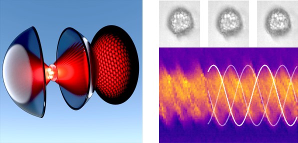
Fig. 1: Novel micromanipulation tools. Left: Holographic optical trap for separate measurements of the forces of each of the laser spots pulling at a trapped cell. Right: Rotation of a biological sample induced by an acoustic wave.
Innovative microscopy: In an FWF-funded collaboration with Prof. Gerhard Schütz’s group at the TU Vienna the ultimate goal is to gain a better understanding of the function of the immune system. The triggering of T-cells on interaction with antigen-presenting cells is investigated. Our group develops optical methods to obtain maximum information (i.e. resolution) from a given number of received photons, whilst minimising the bias effects that are often introduced by experimental boundary conditions, such as optical aberrations.
One recent focus was on the exploitation of near-field effects (supercritical angle fluorescence emission) to improve spatial resolution along the optical axis. The cells are brought into contact with an antigen-functionalised lipid bilayer, which is prepared on a glass coverslip. TIRF illumination restricts the excitation zone to a region that is a few hundred nm thick, which can be resolved in 3D at resolutions down to about 10 nm. Fig. 2 (left) shows an exemplary image of stained microtubules in COS-7 cells. The resolution allows detection of the hollowness of the tubulin structures.
In the Deep brain vision: 3D adaptive 2-photon excitation fluorescence microscopy (FWF project P32146) interdisciplinary project, a Departmental collaboration with the Institute of Physiology (Prof. Michaela Kress), the main research focus on our part is on increasing the imaging depth in scattering media such as brain tissue and on the development of so-called “remote focusing techniques”, which enables quick changing of the focal plane without moving the lens or the specimen, whilst maintaining the highest possible image quality. Fig. 2 (right) shows an image of a GFP-stained microglia cell. The image is a maximum-intensity projection of a recorded 3D stack. Thanks to our sophisticated focusing method, the resolution is independent of the z-position in the focus.
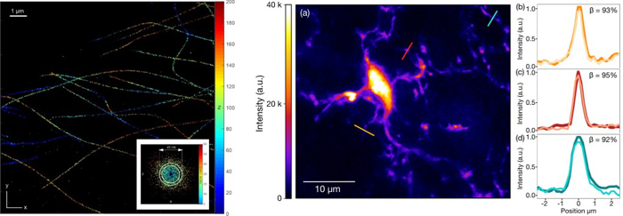
Fig. 2: Innovative microscopy methods. Left: Super-resolution image of (Alexa 647) stained microtubules in fixed COS-7 cells (axial coordinates are colour-coded, units are nm). The inset figure shows an x-z cross-section of a microtubule, constructed by accumulating localisation data along several short strands, revealing its hollow core. Right: Two-photon fluorescence excitation image of a glia cell. The image is an axial maximum-intensity projection over several micrometres. Cross-sections through several processes located at different depths are shown on the right. Dark colour: mechanical z-stepping; light colour: z-stepping using our remote focusing method.
Tunable lenses: The institute also has an international reputation for innovation and expertise in the development of advanced optical elements and optical systems. We have developed (and patented) optical lenses on the basis of a pair of diffractive optical elements (DOEs), so-called Moiré lenses. The focal length of these lenses can be tuned in a wide range by rotating the elements with respect to one another. Recently, the method has been expanded to achieve remote focusing in high numerical aperture microscopes, a highly anticipated new feature in microscopy that was previously impossible (patent pending).
A similar technique was used to design a new zoom system, based solely on rotating DOEs with a quadrupole lens structure – a world first. Typically, DOE-based systems have the problem that they are strongly dispersive, i.e. they are suited only to a certain light colour. In our recent research, however, we have developed a new method to circumvent this issue by using multi-order diffractive optics. This allows us to use these elements in multicoloured and white light implementations, a highly desirable feature for applications in advanced camera systems.
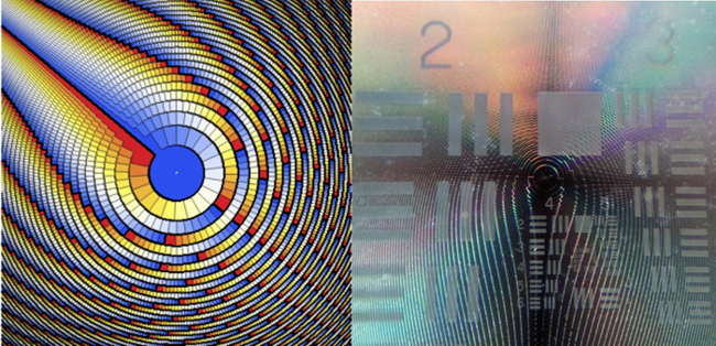
Fig. 3: Tunable lenses. Left: Design pattern of a thin diffractive phase element as part of a focus-tunable diffractive lens operated by rotation. Right: Photo of an accordingly manufactured hardware element.
UV Radiation
Mario Blumthaler, Barbara Klotz, Axel Kreuter, Verena Schenzinger, Michael Schwarzmann and Jochen Wagner
The Solar Radiation and Air Quality research group has been granted an extension of the Austrian UV measuring network project (the UV index, see Fig. 4) for another 10 years, until 2029. As well as continuing routine operations and the calibration of instruments, the project specifically involves the development of a new, state-of-the-art data infrastructure and new analysis, monitoring and visualisation tools. Another focus is on the algorithm development of refined satellite products for UV distribution maps, which also reinforces the group’s collaboration with the European UV research community.
One recent research highlight was the development of a new cloud-screening algorithm for ground-based aerosol remote sensing. The approach is based on a clustering method, as opposed to the traditional thresholding of time series, and it is specifically tailored towards the elimination of thin clouds in aerosol optical depth data. The results are relevant to the major global measuring networks, such as AERONET and GAW, and they demonstrate the expertise of the group within the international aerosol remote sensing community. Furthermore, the group has contributed to the ongoing Austrian project (AMIDA) aimed at developing a prototype data analysis and visualisation platform for the European Space Agency (ESA); a specific usage case for UV data has been tested and documented.
Finally, the group has initiated and is co-leading the Austrian involvement in pan-European ACTRIS (Aerosol, Clouds and Trace Gas Research Infrastructure) activities. In particular, the group laboratory is a designated ACTRIS unit for the calibration of UV-VIS trace gas remote sensing instruments (CREGARS UVVIS-AT). The involvement in ACTRIS is key to future research within the atmospheric science community.
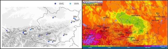
Fig. 4: The institute runs the Austrian UV measuring network, generating the online UV index for Austria, which is updated every few minutes. Left: Austrian UV network measuring stations. Right: Distribution of the UV index over Austria, from ground-based measurements with a satellite-derived cloud attenuation factor.
Selected Publications
- Dholakia, Kishan; Drinkwater, Bruce W.; Ritsch-Marte, Monika: Comparing acoustic and optical forces for biomedical research.
NATURE REVIEW PHYSICS. 2020; 2; 480-491.
https://doi.org/10.1038/s42254-020-0215-3
- Ritsch-Marte, Monika: Orbital angular momentum light in microscopy.
PHILOSOPHICAL TRANSACTIONS OF THE ROYAL SOCIETY A-MATHEMATICAL PHYSICAL AND ENGINEERING SCIENCES. 2017; 375(2087); 20150437.
https://doi.org/10.1098/rsta.2015.0437
- Moser, Simon; Ritsch-Marte, Monika; Thalhammer, Gregor: Model-based compensation of pixel crosstalk in liquid crystal spatial light modulators.
OPTICS EXPRESS. 2019; 27(18); 25046-25063.
https://doi.org/10.1364/OE.27.025046
- May, Molly A.; Bawart, Martin; Langeslag, Michiel; Bernet, Stefan; Kress, Michaela; Ritsch-Marte, Monika; Jesacher, Alexander: High-NA two-photon single cell imaging with remote focusing using a diffractive tunable lens.
BIOMEDICAL OPTICS EXPRESS. 2020; 11(12); 7183-7191.
https://doi.org/10.1364/BOE.405863
- Zelger, Philipp; Bodner, Lisa; Velas, Lukas; Schütz, Gerhard J.; Jesacher, Alexander: Defocused imaging exploits supercritical-angle fluorescence emission for precise axial single molecule localization microscopy.
BIOMEDICAL OPTICS EXPRESS. 2020; 11(2); 775-790.
https://doi.org/10.1364/BOE.375678
- Roider, Clemens; Piestun, Rafael; Jesacher, Alexander: 3D image scanning microscopy with engineered excitation and detection.
OPTICA. 2017; 4(11); 1373-1381.
https://doi.org/10.1364/OPTICA.4.001373
- Bawart, Martin; May, Molly A.; Öttl, Thomas; Roider, Clemens; Bernet, Stefan; Schmidt, Michael; Ritsch-Marte, Monika; Jesacher, Alexander: Diffractive tunable lens for remote focusing in high-NA optical systems.
OPTICS EXPRESS. 2020; 28(18); 26336-26347.
https://doi.org/10.1364/OE.400784
- Bregenzer, Nicola; Öttl, Thomas; Zobernig, Martin; Bawart, Martin; Bernet, Stefan; Ritsch-Marte, Monika: Demonstration of a multi-color diffractive lens with adjustable focal length.
OPTICS EXPRESS. 2020; 28(20); 30150-30163.
https://doi.org/10.1364/OE.404155
- Bernet, Stefan: Zoomable telescope by rotation of toroidal lenses.
APPLIED OPTICS. 2018; 57(27); 8087-8095.
https://doi.org/10.1364/AO.57.008087
Selection of Funding
- FWF: SFB F68 “Tomography Across The Scales”, Teilprojekt “Inverse Problems in Imaging of Trapped Particles” (F 6806-N36), Ritsch-Marte, Monika, € 540k, 2018-2022
- FWF: “3D diffractive elements through fs-laser direct writing” (I 3984-N36) Jesacher Alexander, € 180k, 2020-2023
- FWF: “Deep brain vision: 3D adaptive 2-photon excitation fluorescence microscopy” (P 32146-N36) Jesacher Alexander, € 490k, 2019-2023
- FWF: “3D nanoscopy of the immunological synapse” (P 30214-N36) Jesacher Alexander, Gerhard Schütz, € 350k, 2017-2021
- FWF: “Direct measurement of optical torque” (P 29936-N36) Thalhammer Gregor, € 320k, 2017-2021
- EU: “ACTRIS IMP” Wagner Jochen, € 66k, 2020-2023
- FFG-Bridge: “KombiDOE-Kombinierte diffraktive optische Elemente mit kontinuierlich einstellbaren Eigenschaften“ Bernet Stefan, € 170k, 2018-2021
- FFG: „AMiDA“ Kreuter Axel, € 41k, 2018-2021
- Bundesministerium f. Nachhaltigkeit und Tourismus: „Messung und Analyse der solaren UV Strahlung in Österreich“, Kreuter Axel, € 1.300k, 2019-2029
Collaborations
- Schütz, Gerhard, Technical University of Vienna, Austria
- Schmidt, Michael, Friedrich-Alexander University of Erlangen-Nürnberg, Erlangen, Germany
- Piestun, Rafael, University of Colorado, Boulder, USA
- Wuite, Gijs, Vrije Universiteit Amsterdam, Amsterdam, The Netherlands
- Rubinsztein-Dunlop, Halina, University of Queensland, Brisbane, Australia
- Nevas, Physikalisch-Technische Bundesanstalt, Berlin, Germany
- Gröbner, World Radiation Center, Davos, Switzerland
 o. Univ.-Prof.in Mag.a Dr.in rer.nat. Monika Ritsch-Marte
Director
Contact:
Müllerstraße 44
6020 Innsbruck
Austria
Email: Monika.Ritsch-Marte@i-med.ac.at
Phone: +43 512 9003 70870
Fax: +43 512 9003 73870
www.i-med.ac.at/dpmp/bmp/