


RESPIRATORY CELL PHYSIOLOGY

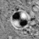
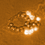
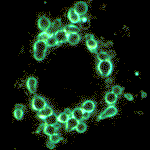 Darkfield microscopy of an AT II cell showing the limiting LB-membranes
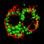
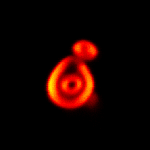
 AT II cells during agonist-induced Ca2+-oscillations
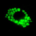
 Polarization microscopy revealing a strong birefringence and a liquid-crystaline structure of intracellular LBs
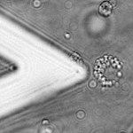
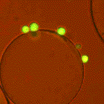
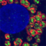
 Stably surface associated LBs (red fluorescent particles) and reflective LB-derived surface structures (cyan)
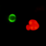
 Vitality of cells judged by retention of a cytosolic marker, Calcein, and absence of nuclear staining by Ethidium homodimer-1 (red)
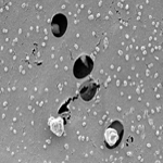
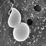
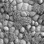 Bronchiolar ciliated cells seen by SEM
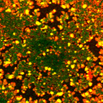
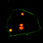
 Co-staining of a type II cell with LTG and Nile Red (yellow). Red clusters mark surfactant in fused vesicles with dissipated proton gradients, green structures acidified endocytotic vesicles and probably kiss-and-run events devoid of lipids
 LBs adsorbed to an air-liquid interface spontaneously transform into lipid expanded (Bodipy-PC; green) and lipid-condensed (DiIC(20); orange) states
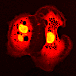
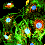 Selective staining of the f-actin cytoskeleton (green), the nucleus (blue) and mitochondria (red) of alveolar type II cells in culture
 Fluorescently labeled Lamellar Bodies (green) of AT II cells are surrounded by highly energized mitochondria (red fluorescence of the mitochondrial potential-sensitive dye JC-1)
 Release of pulmonary surfactant through the exocytotic fusion pore into the extracellular space proceeds by unraveling of the concentrical lipid membranes and their transformation into tubular structures. Surfactant (red) was visualized by FM 1-43, cytosol was stained with Calcein-AM
 The polar tracer Lucifer Yellow preferentially stains cytosol and nucleus of AT II cells leaving out the lumina of Lamellar Bodies (dark inclusions)
 Fusion pore opening of Lamellar Bodies is visualized by FM1-43 (red), which enters the vesicular lumen from the outside and intercalates with lipid surfactant components. Pre-exocytotic vesicles are marked with LTG
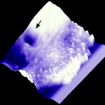 Surface dynamics at the periphery of a stimulated AT II cell observed by atomic force microscopy. Fusion pore opening (arrow) is visible as a deep, transient invagination of the plasma membrane
 DIC microscopy improves the perceptibility of Lamellar Bodies, which appear as large, highly refractive spherical inclusions
 Surface structure of an AT II cell in situ (Image size: 5x5µm)
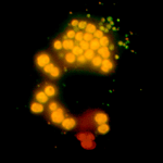
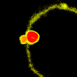
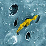 Surfactant protrusion (yellow) out of a fused lamellar body within a cluster of empty fusion pores
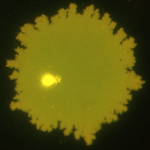
 Freshly released surfactant (center) instantaneously spreads into a surface film (black) when reaching an air-liquid interface
 Surface film (bright area) and remnant structures (dark area, bright spots) after transformation of one lamellar body at a fluorescent air-liquid interface
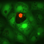
 Type II pneumocyte during global Ca2+-increase (red) and Ca2+-triggered exocytosis (blue spots)
 Surfactant during release through fusion pores usually maintain much of their packed structure (yellow)
 Cantilever of an AFM (the tip cannot be seen) approaching an AT II cell (the central highly granulated one)
 Extracellular surfactant, partially unfolded and unravelled by hydration and separation of the lipid bilayers
 Ballon-like extrusions: The outermost (or innermost) surfactant bilayers frequently separate from the core, suggesting a squeeze-out phenomenon
 AT II cells, microinjected with Lucifer Yellow
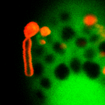
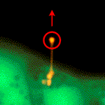 Tearing surfactant out of a fusion pore: Red circle denotes the (invisible) focus of an infrared laser trap
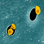
 Living on the edge: Challenging a macrophage with an air-liquid interface
 AT II cells (green) cultured on Solohill Microcarrier Beads
 Confocal z-section of an AT II cell with plasma membrane (yellow) and surfactant (red)
 Interferometry of an AT II cell at an air-liquid interface (bulging of the interface is shown in blue)
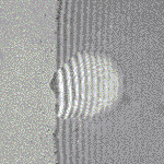 AT II cell at a PFC-water interface (PFC is on the left) and bending of interferometric fringes
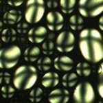
 Lipid organisation at air-liquid interfaces is complex
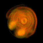 A giant multilamellar vesicle created from organic extracts of native surfactant
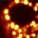
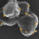
 Distribution of gold nanoparticles (dots) on a surface-adsorbed lamellar body (blue area). Combined darkfield and fluorescence image
 Complex organization of a fluorescent, environment-sensitive styryl dye (FM 1-43) at an inverted air-liquid interface
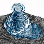
 TEM close-up of internal lamellar body bilayer organization in high pressure/freeze substituted AT II cells
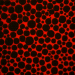
 :Radial symmetric crystals formed out of a melt of DDT. Typical polarization patterns (Maltese cross) are visible
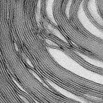
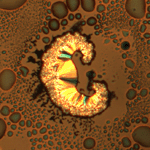
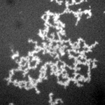 Fractal organization of surface textures by surfactant phospholipids and proteins at an air-liquid interface
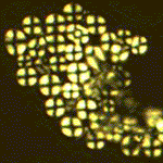
 Near UV fluorescence of Laurdan is revealing a concentrical structure of Intracellular lamellar bodies
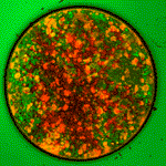
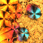
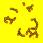
 Starch granules used for optical calibrations in a polarization microscope
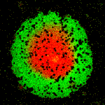
 CARS (Coherent anti-Stokes Raman Spectroscopy) image of an AT II cell demonstrating intracellular lipid deposits
 Separation of the fast (dark) and slow (bright) optical components within a birefringent Lamellar Body in situ (image acquired with crossed polarizers and a retardation plate)
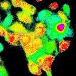
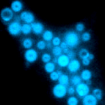
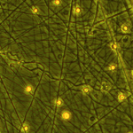
 Primary human AT II cells demonstrating a considerable autofluorescence
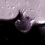
 Gelatine matrix tested as a scaffold in cell culture
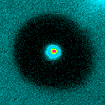
 Unknown object, probably from outer space
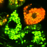
 Immunofluorescence of SP-B (green) and ABCa3 (red) with nuclear stain by Hoechst (blue)
 Immunogold labeling of SP-B (yellow) on extruded pulmonary surfactant, caught in the process of release from exocytosed vesicles in stimulated AT II cells
 Force spectroscopy (Young’s modulus map) with an SP-C functionalized AFM cantilever. Specimen was a Lamellar Body adsorbed on mica
 Z-sectioning of a stimulated cell (plasma membrane: green; apical side: bottom) revealing surfactant (red) containing post-exocytotic vesicles deep inside the cell
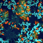
 Following transient fusion and incomplete surfactant release, lamellar bodies are retrieved into the cell (red, FM 1-43), whereas extracellular surfactant is uncovered by alternative dyes (green, SP-DiOC18(3))
 AT II cells cultured at an inverted air-liquid interface (brightfield)
 Generalized Polarization Function (GPF) of Laurdan, sensitive for the extent of water in the polar headgroups of lipid bilayers
 Fractal geometry and low line tension of a surface adsorbed lamellar body
 Fluorescent beads inserted into an inverted air-liquid interface to measure surface dynamics and Marangoni effects
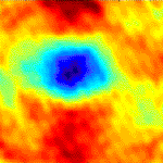
 Calculated homogeneous Ca2+ elevation in a caged Ca2+ and flash-photolysis treated AT II cell
 TEM of an open fusion pore during active squeeze-out of surfactant into the extracellular space
 Giant, surface adsorbed unilamellar vesicle with ongoing lipid phase separation
 Actively degranulating AT II cell seen by DIC and FM 1-43 fluorescence
 Lysotracker Green stain of acidified, intracellular and pre-exocytotic lamellar bodies
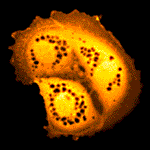
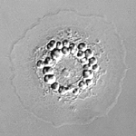
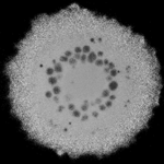
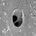 Late stage of compound exocytosis viewed by Scanning Electron Microscopy (SEM)