
 Cellular Electron Microscopy
Our cell biological research concentrates on ultrastructural aspects of membrane trafficking in the context of intracellular signalling, degradation pathways and cellular polarity. Cryo-based (immuno-) electron microscopy and electron tomography are used for studying mammalian cell cultures, human and biopsy material and various eukaryotic model organisms such as mice, yeast, flatworms and Hydra. In addition, we perform methodological research on various organisms from all kingdoms aiming at the improvement and development of specimen and labelling procedures as well as of mammalian cell culture systems.
Selected references
Endosomal signaling and degradation pathways:
Schiefermeier, N., J.M. Scheffler, M.E.G. de Araujo, T. Stasyk, T. Yordaov, H.L. Ebner, M. Offterdinger, S. Munck, M.W. Hess, S.A. Wickström, A. Lange, W. Wunderlich, R. Fässler, D. Teis, and L.A. Huber. 2014 .The late endosomal p14/MP1 (Lamtor2/3) complex regulates focal adhesion dynamics during cell migration. J Cell Biol. in press
Adell, M.A., G.F. Vogel, M. Pakdel, M. Muller, H. Lindner, M.W. Hess, and D. Teis. 2014. Coordinated binding of Vps4 to ESCRT-III drives membrane neck constriction during MVB vesicle formation. J Cell Biol. 205:33-49.
Taub, N., D. Teis, H.L. Ebner, M.W.. 2012. The late endosomal adaptor p14 is a macrophage host-defense factor against Salmonella infection. J Cell Sci. 125:2698-708.
Taub, N., D. Teis, H.L. Ebner, M.W. Hess, and L.A. Huber. 2007. Late endosomal traffic of the epidermal growth factor receptor ensures spatial and temporal fidelity of mitogen-activated protein kinase signaling. Mol Biol Cell. 18:4698-710.
Teis, D., N. Taub, R. Kurzbauer, D. Hilber, M.E. de Araujo, M. Erlacher, M. Offterdinger, A. Villunger, S. Geley, G. Bohn, C. Klein, M.W. Hess, and L.A. Huber. 2006. p14-MP1-MEK1 signaling regulates endosomal traffic and cellular proliferation during tissue homeostasis. J Cell Biol. 175:861-8.
Cellular polarity:
Wiegerinck, C.L., A.R. Janecke, K. Schneeberger, G.F. Vogel, D.Y. van Haaften-Visser, J.C. Escher, R. Adam, C.E. Thoni, K. Pfaller, A.J. Jordan, C.A. Weis, I.J. Nijman, G.R. Monroe, P.M. van Hasselt, E. Cutz, J. Klumperman, H. Clevers, E.E. Nieuwenhuis, R.H. Houwen, G. van Haaften, M.W. Hess, L.A. Huber, J.M. Stapelbroek, T. Müller, and S. Middendorp. 2014. Loss of Syntaxin 3 Causes Variant Microvillus Inclusion Disease. Gastroenterology. in press
Ruemmele, F.M., T. Müller, N. Schiefermeier, H.L. Ebner, S. Lechner, K. Pfaller, C.E. Thoni, O. Goulet, F. Lacaille, J. Schmitz, V. Colomb, F. Sauvat, Y. Revillon, D. Canioni, N. Brousse, G. de Saint-Basile, J. Lefebvre, P. Heinz-Erian, A. Enninger, G. Utermann, M.W. Hess, A.R. Janecke, and L.A. Huber. 2010. Loss-of-function of MYO5B is the main cause of microvillus inclusion disease: 15 novel mutations and a CaCo-2 RNAi cell model. Hum Mutat. 31:544-51.
Muller, T., M.W. Hess, N. Schiefermeier, K. Pfaller, H.L. Ebner, P. Heinz-Erian, H. Ponstingl, J. Partsch, B. Rollinghoff, H. Kohler, T. Berger, H. Lenhartz, B. Schlenck, R.J. Houwen, C.J. Taylor, H. Zoller, S. Lechner, O. Goulet, G. Utermann, F.M. Ruemmele, L.A. Huber, and A.R. Janecke. 2008. MYO5B mutations cause microvillus inclusion disease and disrupt epithelial cell polarity. Nat Genet. 40:1163-5.
Methodology:
Schmiedinger, T., G.F. Vogel, O. Eiter, K. Pfaller, W.A. Kaufmann, A. Flörl, K. Gutleben, S. Schönherr, B. Witting, T.W. Lechleitner, H.L. Ebner, T. Seppi, and M.W. Hess. 2013. Cryo-immunoelectron microscopy of adherent cells improved by the use of electrospun cell culture substrates. Traffic. 14:886-94.
Hess, M.W., K. Pfaller, H.L. Ebner, B. Beer, D. Hekl, and T. Seppi. 2010. 3D versus 2D cell culture implications for electron microscopy. Methods Cell Biol. 96:649-70.
For further publications, see http://scholar.google.com.au/citations?user=pNA_uhoAAAAJ&hl=en
Department für Anatomie, Histologie und Embryologie
 Klinisch angewandte Histologie und Cytologie
- Contact: Guenter.Klima@i-med.ac.at
- Mitarbeiter:
- Ao.Univ.-Prof. Dr. Günter Klima
- Facharzt f.Histologie u.Embryologie, Notarzt
Direktor Division für Histologie und Embryologie
Sprechstunde: Mittwoch 12:00 bis 12:30 - TA Rudolf Haring
langjährige Kooperation mit verschiedenen klinischen Fächern:
- Anaesthesie und allgemeine Intensivmedizin
- Allgemeine Chirurgie
- Kinderchirurgie
- Transplantationschirurgie
- Plastische Chirurgie
- Urologie
- Inst. f. Biochemische Pharmakologie
Diese Zusammenarbeit umfasst einerseits die Cytologische Funktionsüberwachung transplantierter Organe (Fine Needle Aspirationsbiopsie, Pancreatic Juice Cytology, Urinary Sediment Cytology) zur Diagnostik von Abstoßungen, Viralen Infektionen und Medikamententoxischen Funktionseinschränkungen andererseits eine Kooperation bei Großtierversuchen für die Beschaffung, Transport und Betreuung der Versuchstiere (Hausschwein, Mini-Pig, Schaf, Kalb) sowie Intubationsnarkosen bei Kaninchen.
Wesentliche Punkte bei der Organisation von Großtierversuchen sind die richtige Auswahl der Tiere mit einer präoperativen Beobachtung und einem schonenden Transport in sediertem Zustand in einem klimatisierten Transportfahrzeug.
Weiters werden bei Überlebensversuchen von mir der Rücktransport in die Tierversuchsanlage und die postoperative Betreuung (Intensivbetreuung) durchgeführt.
Bei vielen Versuchen wird von meiner Gruppe nach Versuchsbeendigung die histologische Aufarbeitung und Auswertung der Proben unter der fachkundigen Hand von TA Frau Karin Gutleben vorgenommen.
Ein weiteres Aufgabengebiet ist die Planung und Anfertigung von kleinen Laborgeräten (z.B.Rostfreistahlkammer zur Verdauung von Pankreasgewebe zur Inselzellgewinnung)
Für die Lehre wird die Koordination für alte und neue Studienordnung vorgenommen (http://www.uibk.ac.at/c/c5/studien/).
 Working Group for Endothelial Biology
- Contact: Paul.Debbage@i-med.ac.at
phone: +43 (0)512 9003 71178
fax: +43 (0)512 9003 73170
Endothelial biology is at the focus of this group’s research.
Several of our projects are grouped around the biology of blood vessels in tumours (Figures 1, 2).
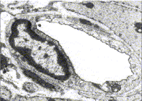 |
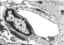
Figure 1: Electron microscope image of a small blood vessel (capillary) |
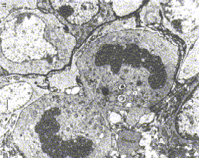 |
Figure 2: Breast carcinoma tumour cell undergoing mitosis (electron microscope image) |
The interactions between endothelial cells and other cell types lie at the heart of the immune response, determine the ability of tumour cells to proliferate and to form metastases, and – last but not least – sculpt the various organs and mediate organ functions.
Pathophysiologies of the vascular system are involved in inflammatory conditions, in tumour growth and spread, and in cardiovascular diseases. Reflecting this, our group collaborates actively with several clinics at the University of Innsbruck, and also internationally with institutes and clinics elsewhere in Europe. In 1998 we presented a technique for intravital lectin perfusion (Debbage et al., 1998) which permits correlative fluorescence and electron microscopical analyses of microvessel density and microvessel permeability (Figures 3,4). In collaboration with the GSF Institutes of Radiobiology (Dr. J. Griebel) and Pathology (Dr. P. Hutzler), and with the Clinic for Radiotherapy and Radio-Oncology (Dr. A. DeVries) at the University of Innsbruck, we showed that fractionated radiotherapy leads to long-standing hyperpermeability of the blood vessels, and also to significant reduction in their density (Figures 3,4). This work formed the basis for successful completion of a doctoral thesis by Dr. Sonja Seidl (2001). Questions arising from that project led to subsequent projects aimed at understanding deeper aspects of endothelial biology.
The group has received funding: Swarovski Foundation, National Bank of Austria, Project 9273.
In the following we introduce here some of our ongoing projects grouped around endothelial cell biology:
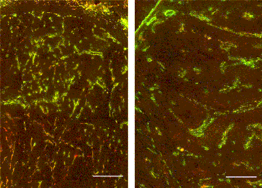 |
| Figure 3,4: Tumour blood vessels, no radiation therapy (3, at left) and after radiation therapy (4, at right) |
1. Endothelial cell biology in tumours: ultrastructural analysis of endothelial cell interactions with prostate tumour cells
- Question:
- How does blood vessel growth relate to metastasis? See Figure 5.
- Collaborations:
-
- Clinic for Urology, (University of Innsbruck), with Professor G. Bartsch
- Institute of Pathology, (University of Innsbruck), with Professor H. Rogatsch
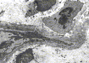 |
Figure 5: Angiogenesis in a prostate carcinoma (electron micrograph, mosaic image) |
 |
|
| Julia Seeber | Elisabeth Wieser |
- Doctoral students carrying out this research:
-
- Miss Elisabeth Wieser (cand. med., Univ. Innsbruck);
- Miss Julia Seeber (cand. med., Univ. Innsbruck).
- Presentations:
-
- At the XVII Congress EAU in Birmingham, March 2002 (Strohmeyer et al., 2002)
- At the 43rd Meeting of the Society for Histochemistry, Vienna, September 2001.
2. Endothelial cell biology in tumours: ultrastructural analysis of endothelial interactions with tumour cells in breast cancers Question: How do the tumour cells penetrate blood vessels during metastasis? See Figure 6.
- Collaborations:
-
- Clinic for General Surgery, (University of Innsbruck) with Dr. M. Dünser
- Clinic for Radiology, (University of Innsbruck) with Professor W. Jaschke and Professor W. Buchberger
- Institute of Pathology (University of Innsbruck with Dr. P. Obrist.
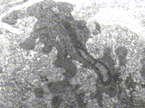 |
Figure 6: A migrating tumour cell in a breast carcinoma (electron microscope image) |
 |
 |
| Silvia Dobler | Silvia Dobler with Paul Debbage |
- Doctoral student carrying out this research:
-
- Miss Silvia Dobler (cand. med., Univ. Innsbruck).
3. Endothelial cell biology in tumours: ultrastructural analysis of radiation effects on blood vessels in a human glioblastoma ectopically implanted in the nude mouse
- Question 1:
- What are the mechanisms of the tumour-endothelial interactions? Tumor-induced growth of small blood vessels is often seen (Figure 7).
- Question 2:
- What are the effects of small radiation doses on the endothelial cells, and on the tumour-endothelial interactions?
- Collaborations:
-
- Department of Radiation Oncology, University Medical Centre Nijmegen, The Netherlands, with Professor A. van der Kogel
- Institute for Pathology of the GSF Research Centre in Munich (Germany),with Dr. P. Hutzler
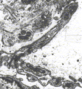 |
Figure 7: Angiogenesis in E106 glioblastoma |
 |
| Eveline Meier |
- Doctoral student carrying out this research:
- Miss Eveline Meier (cand. med., Univ. Innsbruck).
- Presentations:
-
- At the 43rd Meeting of the Society for Histochemistry, Vienna, September 2001
4. Endothelial cell biology in normal tissues: Development of a model system for analysis of radiation effects on human tissues
- Question:
- Can we establish organ-type culture of human tissues which can be used to analyse radiation effects on human blood vessels. Early work on this model began with umbilical vein endothelia (Debbage et al., 2000). Present work focuses on fetal microvessels in the human placental cotyledon (Figure 8).
- Collaborations:
-
- Clinic for Gynaecology and Obstetrics (University of Innsbruck), with Professor C. Marth and Dr. E. Sölder
- Clinic for Radiology I (University of Innsbruck), with Professor W. Jaschke, Professor W. Buchberger and Dr. C. Kremser
- Clinic for Radiotherapy and Radio-Oncology (University of Innsbruck), with Professor P. Lukas
- Institute of Pathology (University of Innsbruck), with Dr. A. Kreczy
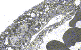 |
Figure 8: Wall of placental villus (electron micrograph) |
- Doctoral student carrying out this research:
-
- Miss Isabella Höliner (cand. med, Univ. Innsbruck).
- Funded by
- the Swarovski Foundation.
- Presentation:
-
- At the 42nd Meeting of the Society for Histochemistry, Les Diablerets, Switzerland.
5. Development of vascular markers for use in Magnetic Resonance Imaging
- Collaborations:
-
- Clinic for Radiology I at the University of Innsbruck,
- Institute for Anorganic Chemistry at the University of Vienna.
 |
 |
| Isabella Höliner with Paul Debbage | Irena Paschkunova |
- Doctoral students carrying out this research:
-
- Miss Isabella Höliner (cand. med, Univ. Innsbruck);
- Miss Irena Paschkunova (Univ. Vienna).
- Funded by
-
- the National Bank of Austria, Project 9273.
Other Work
 |
|
| Karoline Kunz | Klaudia Mistlberger |
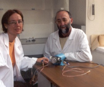
Other work of a technical nature aims to provide new methods of viewing endothelial cells, particularly by use of low voltage scanning electron microscopy. This work is in preparation for publication and formed the basis for the doctoral thesis submitted May 2002 by Miss Karoline Kunz.
Ongoing work is being carried out by Miss Klaudia Mistlberger as part of her doctoral thesis.
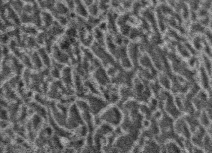 |
Figure 9: LVSEM IMAGE of microvilli |
Occasional collaborations
This group collaborates on an occasional basis with neighbouring research groups at the University of Innsbruck, particularly when the skills available to us complement those of our colleagues in neighbouring institutions. For example, we recently worked with the group of Dr. H. Talasz (Institute for Medical Chemistry, University of Innsbruck) to illustrate the ultrastructure of apoptotic cells (Talasz et al., 2002). We also investigated bone development by immunohistochemical techniques, in collaboration with Professor Dr. H. Fritsch (Institute for Anatomy and Histology, University of Innsbruck) (Fritsch et al., 2001). We collaborated with Professor Michael Hess to demonstrate myelin chemistry in the glacier mummy “Ötzi” (Hess et al., 1998). We have collaborated with Professor Dr. M. Pavelka to visualise endocytotic processes by use of electron histochemical techniques (Hess et al., 2000; Pavelka et al., 1998).
Technical colleagues
 |
| Julianna Forgo |
- Mrs. Julianna Forgo,
- Mr. Rudolf Haring.
Selected citations (primary literature)
- Debbage, Pl; Griebel, J; Ried, M; Gneiting, T; DeVries, A; Hutzler, P (1998) Lectin intravital perfusion studies in tumor-bearing mice: micrometer-resolution, wide-area mapping of microvascular labeling, distinguishing efficiently and inefficiently perfused microregions in the tumor. J Histochem Cytochem. 46:627-39
- Debbage PL, Seidl S, Kreczy A, Hutzler P, Pavelka M & Lukas P (2000) Vascular permeability and hyperpermeability in a murine adenocarcinoma after fractionated radiotherapy: an ultrastructural tracer study. Histochem. Cell Biol. 114: 259-275
- Debbage PL, Sölder E, Seidl S, Hutzler P, Hugl B, Öfner D & Kreczy A (2001) Intravital lectin perfusion analysis of vascular permeability in human micro- and macro- blood vessels. Histochem. Cell Biol. 116: 349-359
- Fritsch H, Brenner E & Debbage P (2001) Ossification in the human calcaneus: a model for spatial bone development and ossification. J Anat. 199: 609-16
- Hess MW, Kirschning E, Pfaller K, Debbage Pl, Hohenberg H & Klima G (1998) 5000-year-old myelin: uniquely intact in molecular configuration and fine structure. Curr. Biol. 8: R512-3
- Hess MW, Muller M, Debbage Pl, Vetterlein M & Pavelka M (2000( Cryopreparation provides new insight into effects of brefeldin ! on the structure of the HepG2 Golgi apparatus. J. Struct. Biol. 130: 63-72
- Pavelka M, Ellinger A, Debbage P, Loewe C, Vetterlein M & Roth J (1998) Endocytic routes to the Golgi apparatus. Histochem. Cell Biol. 109: 555-70
- Strohmeyer et al. (2002) Birmingham meeting xxxxxxxxx
- Talasz H, Helliger W, Sarg B, Debbage PL, Puschendorf B & Lindner H (2002) Hyperphosphorylation of histone H2A.X and dephosphorylation of histone H1 subtypes in the course of apoptosis. Cell Death Differ. 9: 27-39
Doctoral theses
- Seidl S (2001) Doctoral thesis, Medical Faculty of the University of Innsbruck: Vaskuläre Veränderungen in AT17-Tumoren der Maus während Strahlentherapie
- Kunz K (2002) Doctoral thesis, Medical Faculty of the University of Innsbruck: Low voltage scanning electron microscopic analysis of epithelial cell surfaces: characterisation of a carbohydrate rich surface layer.
Contact
Michael W. Hess, PhD
Associate Professor of Cell Biology
telephone: +43 (0)512 9003 71176
Michael.Hess@i-med.ac.at
Georg F. Vogel, MD
University Assistant
telephone:+ 43 (0)512 9003 71181
Georg.Vogel@i-med.ac.at
Karin Gutleben
Research Technician
telephone: +43 (0)512 9003 71177
Karin.Gutleben@i-med.ac.at
Barbara Witting, BMA
Research Technician
telephone: +43 (0)512 9003 71177
Barbara.Witting@i-med.ac.at
Klinisch angewandte Histologie und Cytologie
- Contact: Guenter.Klima@i-med.ac.at
- Mitarbeiter:
- Ao.Univ.-Prof. Dr. Günter Klima
- Facharzt f.Histologie u.Embryologie, Notarzt
Direktor Division für Histologie und Embryologie
Sprechstunde: Mittwoch 12:00 bis 12:30 - TA Rudolf Haring
langjährige Kooperation mit verschiedenen klinischen Fächern:
- Anaesthesie und allgemeine Intensivmedizin
- Allgemeine Chirurgie
- Kinderchirurgie
- Transplantationschirurgie
- Plastische Chirurgie
- Urologie
- Inst. f. Biochemische Pharmakologie
Diese Zusammenarbeit umfasst einerseits die Cytologische Funktionsüberwachung transplantierter Organe (Fine Needle Aspirationsbiopsie, Pancreatic Juice Cytology, Urinary Sediment Cytology) zur Diagnostik von Abstoßungen, Viralen Infektionen und Medikamententoxischen Funktionseinschränkungen andererseits eine Kooperation bei Großtierversuchen für die Beschaffung, Transport und Betreuung der Versuchstiere (Hausschwein, Mini-Pig, Schaf, Kalb) sowie Intubationsnarkosen bei Kaninchen.
Wesentliche Punkte bei der Organisation von Großtierversuchen sind die richtige Auswahl der Tiere mit einer präoperativen Beobachtung und einem schonenden Transport in sediertem Zustand in einem klimatisierten Transportfahrzeug.
Weiters werden bei Überlebensversuchen von mir der Rücktransport in die Tierversuchsanlage und die postoperative Betreuung (Intensivbetreuung) durchgeführt.
Bei vielen Versuchen wird von meiner Gruppe nach Versuchsbeendigung die histologische Aufarbeitung und Auswertung der Proben unter der fachkundigen Hand von TA Frau Karin Gutleben vorgenommen.
Ein weiteres Aufgabengebiet ist die Planung und Anfertigung von kleinen Laborgeräten (z.B.Rostfreistahlkammer zur Verdauung von Pankreasgewebe zur Inselzellgewinnung)
Für die Lehre wird die Koordination für alte und neue Studienordnung vorgenommen (http://www.uibk.ac.at/c/c5/studien/).
Working Group for Endothelial Biology
- Contact: Paul.Debbage@i-med.ac.at
phone: +43 (0)512 9003 71178
fax: +43 (0)512 9003 73170
Endothelial biology is at the focus of this group’s research.
Several of our projects are grouped around the biology of blood vessels in tumours (Figures 1, 2).
|
Figure 1: Electron microscope image of a small blood vessel (capillary) |
|
Figure 2: Breast carcinoma tumour cell undergoing mitosis (electron microscope image) |
The interactions between endothelial cells and other cell types lie at the heart of the immune response, determine the ability of tumour cells to proliferate and to form metastases, and – last but not least – sculpt the various organs and mediate organ functions.
Pathophysiologies of the vascular system are involved in inflammatory conditions, in tumour growth and spread, and in cardiovascular diseases. Reflecting this, our group collaborates actively with several clinics at the University of Innsbruck, and also internationally with institutes and clinics elsewhere in Europe. In 1998 we presented a technique for intravital lectin perfusion (Debbage et al., 1998) which permits correlative fluorescence and electron microscopical analyses of microvessel density and microvessel permeability (Figures 3,4). In collaboration with the GSF Institutes of Radiobiology (Dr. J. Griebel) and Pathology (Dr. P. Hutzler), and with the Clinic for Radiotherapy and Radio-Oncology (Dr. A. DeVries) at the University of Innsbruck, we showed that fractionated radiotherapy leads to long-standing hyperpermeability of the blood vessels, and also to significant reduction in their density (Figures 3,4). This work formed the basis for successful completion of a doctoral thesis by Dr. Sonja Seidl (2001). Questions arising from that project led to subsequent projects aimed at understanding deeper aspects of endothelial biology.
The group has received funding: Swarovski Foundation, National Bank of Austria, Project 9273.
In the following we introduce here some of our ongoing projects grouped around endothelial cell biology:
|
| Figure 3,4: Tumour blood vessels, no radiation therapy (3, at left) and after radiation therapy (4, at right) |
1. Endothelial cell biology in tumours: ultrastructural analysis of endothelial cell interactions with prostate tumour cells
- Question:
- How does blood vessel growth relate to metastasis? See Figure 5.
- Collaborations:
-
- Clinic for Urology, (University of Innsbruck), with Professor G. Bartsch
- Institute of Pathology, (University of Innsbruck), with Professor H. Rogatsch
|
Figure 5: Angiogenesis in a prostate carcinoma (electron micrograph, mosaic image) |
|
|
| Julia Seeber | Elisabeth Wieser |
- Doctoral students carrying out this research:
-
- Miss Elisabeth Wieser (cand. med., Univ. Innsbruck);
- Miss Julia Seeber (cand. med., Univ. Innsbruck).
- Presentations:
-
- At the XVII Congress EAU in Birmingham, March 2002 (Strohmeyer et al., 2002)
- At the 43rd Meeting of the Society for Histochemistry, Vienna, September 2001.
2. Endothelial cell biology in tumours: ultrastructural analysis of endothelial interactions with tumour cells in breast cancers Question: How do the tumour cells penetrate blood vessels during metastasis? See Figure 6.
- Collaborations:
-
- Clinic for General Surgery, (University of Innsbruck) with Dr. M. Dünser
- Clinic for Radiology, (University of Innsbruck) with Professor W. Jaschke and Professor W. Buchberger
- Institute of Pathology (University of Innsbruck with Dr. P. Obrist.
|
Figure 6: A migrating tumour cell in a breast carcinoma (electron microscope image) |
|
|
| Silvia Dobler | Silvia Dobler with Paul Debbage |
- Doctoral student carrying out this research:
-
- Miss Silvia Dobler (cand. med., Univ. Innsbruck).
3. Endothelial cell biology in tumours: ultrastructural analysis of radiation effects on blood vessels in a human glioblastoma ectopically implanted in the nude mouse
- Question 1:
- What are the mechanisms of the tumour-endothelial interactions? Tumor-induced growth of small blood vessels is often seen (Figure 7).
- Question 2:
- What are the effects of small radiation doses on the endothelial cells, and on the tumour-endothelial interactions?
- Collaborations:
-
- Department of Radiation Oncology, University Medical Centre Nijmegen, The Netherlands, with Professor A. van der Kogel
- Institute for Pathology of the GSF Research Centre in Munich (Germany),with Dr. P. Hutzler
| |
Figure 7: Angiogenesis in E106 glioblastoma |
|
| Eveline Meier |
- Doctoral student carrying out this research:
- Miss Eveline Meier (cand. med., Univ. Innsbruck).
- Presentations:
-
- At the 43rd Meeting of the Society for Histochemistry, Vienna, September 2001
4. Endothelial cell biology in normal tissues: Development of a model system for analysis of radiation effects on human tissues
- Question:
- Can we establish organ-type culture of human tissues which can be used to analyse radiation effects on human blood vessels. Early work on this model began with umbilical vein endothelia (Debbage et al., 2000). Present work focuses on fetal microvessels in the human placental cotyledon (Figure 8).
- Collaborations:
-
- Clinic for Gynaecology and Obstetrics (University of Innsbruck), with Professor C. Marth and Dr. E. Sölder
- Clinic for Radiology I (University of Innsbruck), with Professor W. Jaschke, Professor W. Buchberger and Dr. C. Kremser
- Clinic for Radiotherapy and Radio-Oncology (University of Innsbruck), with Professor P. Lukas
- Institute of Pathology (University of Innsbruck), with Dr. A. Kreczy
|
Figure 8: Wall of placental villus (electron micrograph) |
- Doctoral student carrying out this research:
-
- Miss Isabella Höliner (cand. med, Univ. Innsbruck).
- Funded by
- the Swarovski Foundation.
- Presentation:
-
- At the 42nd Meeting of the Society for Histochemistry, Les Diablerets, Switzerland.
5. Development of vascular markers for use in Magnetic Resonance Imaging
- Collaborations:
-
- Clinic for Radiology I at the University of Innsbruck,
- Institute for Anorganic Chemistry at the University of Vienna.
|
|
| Isabella Höliner with Paul Debbage | Irena Paschkunova |
- Doctoral students carrying out this research:
-
- Miss Isabella Höliner (cand. med, Univ. Innsbruck);
- Miss Irena Paschkunova (Univ. Vienna).
- Funded by
-
- the National Bank of Austria, Project 9273.
Other Work
|
|
| Karoline Kunz | Klaudia Mistlberger |
Other work of a technical nature aims to provide new methods of viewing endothelial cells, particularly by use of low voltage scanning electron microscopy. This work is in preparation for publication and formed the basis for the doctoral thesis submitted May 2002 by Miss Karoline Kunz.
Ongoing work is being carried out by Miss Klaudia Mistlberger as part of her doctoral thesis.
|
Figure 9: LVSEM IMAGE of microvilli |
Occasional collaborations
This group collaborates on an occasional basis with neighbouring research groups at the University of Innsbruck, particularly when the skills available to us complement those of our colleagues in neighbouring institutions. For example, we recently worked with the group of Dr. H. Talasz (Institute for Medical Chemistry, University of Innsbruck) to illustrate the ultrastructure of apoptotic cells (Talasz et al., 2002). We also investigated bone development by immunohistochemical techniques, in collaboration with Professor Dr. H. Fritsch (Institute for Anatomy and Histology, University of Innsbruck) (Fritsch et al., 2001). We collaborated with Professor Michael Hess to demonstrate myelin chemistry in the glacier mummy “Ötzi” (Hess et al., 1998). We have collaborated with Professor Dr. M. Pavelka to visualise endocytotic processes by use of electron histochemical techniques (Hess et al., 2000; Pavelka et al., 1998).
Technical colleagues
|
| Julianna Forgo |
- Mrs. Julianna Forgo,
- Mr. Rudolf Haring.
Selected citations (primary literature)
- Debbage, Pl; Griebel, J; Ried, M; Gneiting, T; DeVries, A; Hutzler, P (1998) Lectin intravital perfusion studies in tumor-bearing mice: micrometer-resolution, wide-area mapping of microvascular labeling, distinguishing efficiently and inefficiently perfused microregions in the tumor. J Histochem Cytochem. 46:627-39
- Debbage PL, Seidl S, Kreczy A, Hutzler P, Pavelka M & Lukas P (2000) Vascular permeability and hyperpermeability in a murine adenocarcinoma after fractionated radiotherapy: an ultrastructural tracer study. Histochem. Cell Biol. 114: 259-275
- Debbage PL, Sölder E, Seidl S, Hutzler P, Hugl B, Öfner D & Kreczy A (2001) Intravital lectin perfusion analysis of vascular permeability in human micro- and macro- blood vessels. Histochem. Cell Biol. 116: 349-359
- Fritsch H, Brenner E & Debbage P (2001) Ossification in the human calcaneus: a model for spatial bone development and ossification. J Anat. 199: 609-16
- Hess MW, Kirschning E, Pfaller K, Debbage Pl, Hohenberg H & Klima G (1998) 5000-year-old myelin: uniquely intact in molecular configuration and fine structure. Curr. Biol. 8: R512-3
- Hess MW, Muller M, Debbage Pl, Vetterlein M & Pavelka M (2000( Cryopreparation provides new insight into effects of brefeldin ! on the structure of the HepG2 Golgi apparatus. J. Struct. Biol. 130: 63-72
- Pavelka M, Ellinger A, Debbage P, Loewe C, Vetterlein M & Roth J (1998) Endocytic routes to the Golgi apparatus. Histochem. Cell Biol. 109: 555-70
- Strohmeyer et al. (2002) Birmingham meeting xxxxxxxxx
- Talasz H, Helliger W, Sarg B, Debbage PL, Puschendorf B & Lindner H (2002) Hyperphosphorylation of histone H2A.X and dephosphorylation of histone H1 subtypes in the course of apoptosis. Cell Death Differ. 9: 27-39
Doctoral theses
- Seidl S (2001) Doctoral thesis, Medical Faculty of the University of Innsbruck: Vaskuläre Veränderungen in AT17-Tumoren der Maus während Strahlentherapie
- Kunz K (2002) Doctoral thesis, Medical Faculty of the University of Innsbruck: Low voltage scanning electron microscopic analysis of epithelial cell surfaces: characterisation of a carbohydrate rich surface layer.
Contact
Michael W. Hess, PhD
Associate Professor of Cell Biology
telephone: +43 (0)512 9003 71176
Michael.Hess@i-med.ac.at
Georg F. Vogel, MD
University Assistant
telephone:+ 43 (0)512 9003 71181
Georg.Vogel@i-med.ac.at
Karin Gutleben
Research Technician
telephone: +43 (0)512 9003 71177
Karin.Gutleben@i-med.ac.at
Barbara Witting, BMA
Research Technician
telephone: +43 (0)512 9003 71177
Barbara.Witting@i-med.ac.at